|
|
|
| Is fluoroscopy-free single-use flexible ureteroscopy a feasible treatment for kidney stones with abnormal renal anatomy? |
Hamdy Aboutaleba,Mohamed Sultana,Ahmed Zaghloulb,Yasser Farahatb,Maher Gawishc,Fouad Zanatya,*( ) )
|
aUrology Department, Menoufia University Hospital, Egypt
bUrology Department, Burjeel Hospital, Abu Dhabi, United Arab Emirates
cUrology Department, AlAzhar University, Assiut Branch, Egypt |
|
|
|
|
Abstract Objective: This study aimed to evaluate the feasibility of the fluoroscopy-free single-use flexible ureteroscopy procedure in the treatment of kidney stones with abnormal renal anatomy compared to normal renal anatomy. Methods: Forty patients with abnormal (Group A) and 80 patients with normal (Group B) renal anatomy who had 10-20 mm renal stones were included. They were treated with LithoVue single-use flexible ureteroscopy (Boston Scientific, Marlborough, MA, USA) after ureteric dilatation by two different size semi-rigid ureteroscopes. This technique was chosen as the aim was to exclude any ureteric pathology (e.g., stone or stricture), confirm the placement of a safe guidewire, avoid balloon dilatation of the ureter, and achieve safe insertion of a 12 Fr, 35/45 cm ureteric access sheath with optical and tactile sign and without fluoroscopy image for guidance. Results: The mean ages were 43 years and 45 years in Group A and Group B, respectively. The mean stone burden was 14.62 (standard deviation: 5.35) mm3 and 14.79 (standard deviation: 4.58) mm3 in Group A and Group B, respectively. There is no significant difference between both groups according to the mean operative time, hospital stay, or stone-free rate. The stone-free rate was about 93% in both groups when the stone size was between 10 mm and 15 mm, and less than 54% when the stone size was more than 15 mm to 20 mm. In the majority of cases (80.0% in Group A and 92.5% in Group B), we completed the procedure without fluoroscopy. The perioperative complication rates were comparable in the two groups. Conclusion: Fluoroscopy-free single-use flexible ureteroscopy, when performed by expert urologists, is a feasible treatment for pre-stented patients with kidney calculi of ≤15 mm with abnormal renal anatomy.
|
|
Received: 24 January 2023
Available online: 20 October 2024
|
|
Corresponding Authors:
* E-mail address: drfouad80@gmail.com (F. Zanaty).
|
|
|
| Demographic data and stone characteristics | Group Aa (n=40) | Group Bb (n=80) | p-Value | | Age, year | | | 0.62 | | Mean±SD | 43±14 | 45±13 | | Median (IQR) | 45 (29-57) | 43 (32-58) | | Sexc | | | 0.87 | | Male | 32 (80.0) | 65 (81.2) | | Female | 8 (20.0) | 15 (18.8) | | Kidney sidec | | | 0.57 | | Right kidney | 10 (25.0) | 24 (30.0) | | Left kidney | 30 (75.0) | 56 (70.0) | | Clinical presentationc | | | 0.49 | | Symptomatic | 32 (80.0) | 68 (85.0) | | Asymptomatic | 8 (20.0) | 12 (15.0) | | Stone burden in CT scan, mm3 | 0.69 | | Mean±SD | 14.62±5.35 | 14.79±4.58 | | Median (IQR) | 12.3 (6.0-20.0) | 12.4 (6.0-20.0) | | Stone density, HU | | | 0.002 | | Mean±SD | 1132.3±244.5 | 977.5±311.6 | | Median (IQR) | 830 (714-1517) | 821 (460-1744) | | Stone locationc | | Pelvis | 14 (35.0) | 26 (32.5) | 0.54 | | Lower | 9 (22.5) | 18 (22.5) | 0.82 | | Middle | 4 (10.0) | 8 (10.0) | 0.56 | | Upper | 3 (7.5) | 6 (7.5) | 0.33 | | Multiple | 10 (25.0) | 22 (27.5) | 0.33 | | Stone numberc | | | 0.33 | | Single | 30 (75.0) | 58 (72.5) | | Multiple | 10 (25.0) | 22 (27.5) | | Stone burdenc | | | 0.42 | | 10-15 mm3 | 28 (70.0) | 50 (62.5) | | >15-20 mm3 | 12 (30.0) | 30 (37.5) |
|
|
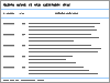
Demographic data and stone characteristics of the studied groups.
|
| Variable | Group Aa (n=40) | Group Bb (n=80) | p-Value | | Operative time, min | | Mean±SD | 68.68±28.4 | 64.37±22.28 | 0.31 | | Median (IQR) | 60 (30-108) | 58 (35-99) | 0.29 | | Need for DJ ureteric stent, n (%) | 35 (87.5) | 50 (62.5) | 0.42 | | UAS insertion, n (%) | 19 (47.5) | 74 (92.5) | 0.01c | | Use of fluoroscopy guidance, n (%) | 8 (20.0) | 6 (7.5) | 0.04c | | Hospital stay, h | | | 0.72 | | Mean±SD | 17.85±4.94 | 19.3±7.72 | | Median (IQR) | 10 (6-24) | 11 (8-32) | | Stone-free rate, n/N (%) | | Stone burden (10-15 mm3) | 26/28 (92.9) | 46/50 (92.0) | 0.71 | | Stone burden (>15-20 mm3) | 6/12 (50.0) | 16/30 (53.3) | 0.17 | | Lower calyx | 6/9 (66.7) | 16/18 (88.9) | 0.38 | | Postoperative complication, n (%) | | Postoperative hematuria (Grade IId) | 5 (12.5) | 9 (11.2) | 0.62 | | Ureteric wall injury (Grade Id) | 3 (7.5) | 5 (6.2) | 0.22 | | UTI (Grade IId) | 2 (5.0) | 4 (5.0) | 0.85 |
|
|
Operative and postoperative data among the studied groups.
|
| [1] |
Raj GV, Auge BK, Assimos D, Preminger GM. Metabolic abnormalities associated with renal calculi in patients with horseshoe kidneys. J Endourol 2004; 18:157-61.
doi: 10.1089/089277904322959798
pmid: 15072623
|
| [2] |
Giusti G, Proietti S, Peschechera R, Taverna G, Sortino G, Cindolo L, et al. Sky is no limit for ureteroscopy: extending the indications and special circumstances. World J Urol 2015; 33:257-73.
doi: 10.1007/s00345-014-1345-y
pmid: 24962930
|
| [3] |
Berrington de González A, Darby S. Risk of cancer from diagnostic X-rays: estimates for the UK and 14 other countries. Lancet 2004; 363:345-51.
doi: 10.1016/S0140-6736(04)15433-0
pmid: 15070562
|
| [4] |
Manzo BO, Lozada E, Manzo G, Sánchez HM, Gomez F, Figueroa A, et al. Radiation-free flexible ureteroscopy for kidney stone treatment. Arab J Urol 2019; 17:200-5.
|
| [5] |
Guven S, Yigit P, Tuncel A, Karabulut I, Sahin S, Kilic O, et al. Retrograde intrarenal surgery of renal stones: a critical multiaspect evaluation of the outcomes by the Turkish Academy of Urology Prospective Study Group (ACUP Study). World J Urol 2021; 39:549-54.
|
| [6] |
Türk C, Pet?ík A, Sarica K, Seitz C, Skolarikos A, Straub M, et al. EAU guidelines on interventional treatment for urolithiasis. Eur Urol 2016; 69:475-82.
doi: 10.1016/j.eururo.2015.07.041
pmid: 26344917
|
| [7] |
Aboutaleb H. Fluoroscopy free flexible ureteroscopy with holmium:yttrium-aluminium-garnet laser lithotripsy for removal of renal calculi. Arab J Urol 2016; 14:123-30.
|
| [8] |
Dindo D, Demartines N, Clavien PA. Classification of surgical complications: a new proposal with evaluation in a cohort of 6336 patients and results of a survey. Ann Surg 2004; 240:205-13.
doi: 10.1097/01.sla.0000133083.54934.ae
pmid: 15273542
|
| [9] |
Schoenthaler M, Buchholz N, Farin E, Ather H, Bach C, Bach T, et al. The Post-Ureteroscopic Lesion Scale (PULS): a multicenter video-based evaluation of inter-rater reliability. World J Urol 2014; 32:1033-40.
doi: 10.1007/s00345-013-1185-1
pmid: 24135917
|
| [10] |
Stein RJ, Desai MM. Management of urolithiasis in the congenitally abnormal kidney (horseshoe and ectopic). Curr Opin Urol 2007; 17:125-31.
pmid: 17285023
|
| [11] |
Theiss M, Hofmockel G, Fromüller H. [Laparoscopy in urology. Current status and perspectives]. Dtsch Med Wochenschr 1993; 118:1331-5. [Article in German].
|
| [12] |
Tunc L, Tokgoz H, Tan MO, Kupeli B, Karaoglan U, Bozkirli I. Stones in anomalous kidneys: results of treatment by shock wave lithotripsy in 150 patients. Int J Urol 2004; 11:831-6.
pmid: 15479286
|
| [13] |
Ganpule AP, Desai MR. Urolithiasis in kidneys with abnormal lie, rotation or form. Curr Opin Urol 2011; 21:145-53.
doi: 10.1097/MOU.0b013e3283435c79
pmid: 21285720
|
| [14] |
Astolfi RH, Freschi G, Berti FF, Gattas N, Molina WRJ, Meller A. Flexible ureterorenoscopy in position or fusion anomaly: is it feasible? Rev Assoc Med Bras 2017; 63:685-8.
|
| [15] |
Kirac M, Kopru B, Ergin G, Kibar Y, Biri H. Is fluoroscopy necessary during flexible ureteroscopy for the treatment of renal stones? Arab J Urol 2019; 18:112-7.
|
| [16] |
Giblin JG, Rubenstein J, Taylor A, Pahira J. Radiation risk to the urologist during endourologic procedures, and a new shield that reduces exposure. Urology 1996; 48:624-7.
pmid: 8886071
|
| [17] |
Chen TT, Wang C, Ferrandino MN, Scales CD, Yoshizumi TT, Preminger GM, et al. Radiation exposure during the evaluation and management of nephrolithiasis. J Urol 2015; 194:878-85.
doi: 10.1016/j.juro.2015.04.118
pmid: 26055822
|
| [18] |
Olgin G, Smith D, Alsyouf M, Arenas JL, Engebretsen S, Huang G, et al. Ureteroscopy without fluoroscopy: a feasibility study and comparison with conventional ureteroscopy. J Endourol 2015; 29:625-9.
doi: 10.1089/end.2014.0237
pmid: 25562139
|
| [19] |
Emiliani E, Kanashiro A, Chi T, Pérez-Fentes DA, Manzo BO, Angerri O, et al. Fluoroless endourological surgery for stone disease: a review of the literature-tips and tricks. Curr Urol Rep 2020; 21:27. https://doi.org/10.1007/s11934-020-00979-y.
doi: 10.1007/s11934-020-00979-y
pmid: 32444987
|
| [20] |
Majdalany SE, Levin BA, Ghani KR. The efficiency of Moses technology holmium laser for treating renal stones during flexible ureteroscopy: relationship between stone volume, time, and energy. J Endourol 2021; 35(Suppl. 3):S14-21. https://doi.org/10.1089/end.2021.0592.
doi: 10.1089/end.2021.0592
pmid: 34910609
|
| [21] |
Aldoukhi AH, Black KM, Ghani KR. Emerging laser techniques for the management of stones. Urol Clin 2019; 46:193-205.
|
| [22] |
Grasso M, Conlin M, Bagley D. Retrograde ureteropyeloscopic treatment of 2 cm or greater upper urinary tract and minor staghorn calculi. J Urol 1998; 160:346-51.
pmid: 9679874
|
| [23] |
Portis AJ, Rygwall R, Holtz C, Pshon N, Laliberte M. Ureteroscopic laser lithotripsy for upper urinary tract calculi with active fragment extraction and computerized tomography followup. J Urol 2006; 175:2129-34.
doi: 10.1016/S0022-5347(06)00311-9
pmid: 16697818
|
| [24] |
Takazawa R, Kitayama S, Tsujii T. Successful outcome of flexible ureteroscopy with holmium laser lithotripsy for renal stones 2 cm or greater. Int J Urol 2012; 19:264-7.
doi: 10.1111/j.1442-2042.2011.02931.x
pmid: 22145599
|
| [25] |
Lavan L, Herrmann T, Netsch C, Becker B, Somani BK. Outcomes of ureteroscopy for stone disease in anomalous kidneys: a systematic review. World J Urol 2020; 38:1135-46.
doi: 10.1007/s00345-019-02810-x
pmid: 31101967
|
| [26] |
Mahmood SN, Toffeq H, Fakhralddin S. Sheathless and fluoroscopy-free retrograde intrarenal surgery: an attractive way of renal stone management in high-volume stone centers. Asian J Urol 2020; 7:309-17.
doi: 10.1016/j.ajur.2019.07.003
pmid: 32742931
|
| [27] |
Senel C, Tuncel A, Balci M, Asfuroglu A, Aykanat C, Guzel O, et al. Safety and reliability of fluoroscopy-free technique in retrograde intrarenal surgery. Minerva Urol Nefrol 2018; 70:606-11.
doi: 10.23736/S0393-2249.18.03228-9
pmid: 30230298
|
| [1] |
Makoto Taguchi, Kaneki Yasuda, Hidefumi Kinoshita. The optimal stent pusher position to achieve successful ureteral stent insertion under fluoroscopic guidance[J]. Asian Journal of Urology, 2024, 11(2): 311-315. |
| [2] |
Qinghui Wu,Kesavan Esuvaranathan,Teck Kheng Lee,Soo Leong Foo,Jian Ping Chai,Edmund Chiong. A pilot clinical study of developing an External Assist Targeting Device for rapid and precise renal calyx access during percutaneous nephrolithotomy[J]. Asian Journal of Urology, 2023, 10(3): 364-371. |
| [3] |
Yuanshen Mao,Wenfeng Li,Jun Da,Mingxi Xu,Yiwei Wang,Yufei Gu,Weixin Pan,Zhong Wang. Analysis of the effect of holmium laser flexible ureteroscopic intrapelvic drainage in the treatment of parapelvic renal cysts[J]. Asian Journal of Urology, 2023, 10(2): 172-176. |
| [4] |
. Reliability of nephrolithometric nomograms in patients treated with minimally invasive percutaneous nephrolithotomy: A precision study[J]. Asian Journal of Urology, 2023, 10(1): 70-80. |
| [5] |
Sujeet Poudyal. Current insights on haemorrhagic complications in percutaneous nephrolithotomy[J]. Asian Journal of Urology, 2022, 9(1): 81-93. |
| [6] |
Dilip K. Mishra,Sonia Bhatt,Sundaram Palaniappan,Talamanchi V.K. Reddy,Vinothkumar Rajenthiran,Y.L. Sreeranga,Madhu S. Agrawal. Mini versus ultra-mini percutaneous nephrolithotomy in a paediatric population[J]. Asian Journal of Urology, 2022, 9(1): 75-80. |
| [7] |
Braulio Omar Manzo,Jose David Cabrera,Esteban Emiliani,Hector Manuel Sánchez,Brian Howard Eisner,Jose Ernesto Torres. Impact of the adherence to medical treatment on the main urinary metabolic disorders in patients with kidney stones[J]. Asian Journal of Urology, 2021, 8(3): 275-279. |
| [8] |
Sarwar Noori Mahmood,Hewa Toffeq,Saman Fakhralddin. Sheathless and fluoroscopy-free retrograde intrarenal surgery: An attractive way of renal stone management in high-volume stone centers[J]. Asian Journal of Urology, 2020, 7(3): 309-317. |
| [9] |
Aso Omer Rashid, Saman Salih Fakhulddin. Risk factors for fever and sepsis after percutaneous nephrolithotomy[J]. Asian Journal of Urology, 2016, 3(2): 82-87. |
| [10] |
Yasser A. Noureldin, Mohamed A. Elkoushy, Sero Andonian. Does the presence of a percutaneous renal access influence fluoroscopy time during percutaneous nephrolithotomy?[J]. Asian Journal of Urology, 2015, 2(4): 220-223. |
| [11] |
Husain Alenezi, John D. Denstedt. Flexible ureteroscopy: Technological advancements, current indications and outcomes in the treatment of urolithiasis[J]. Asian Journal of Urology, 2015, 2(3): 133-141. |
|
|
|
|