Three-dimensional automatic artificial intelligence driven augmented-reality selective biopsy during nerve-sparing robot-assisted radical prostatectomy: A feasibility and accuracy study
Enrico Checcuccia *(),Alberto Pianab,Gabriele Volpia,Pietro Piazzollac,Daniele Amparoreb,Sabrina De Cillisb,Federico Piramideb,Cecilia Gattib,Ilaria Sturad,Enrico Bollitoe,Federica Massae,Michele Di Diof,Cristian Fiorib,Francesco Porpigliab
aDepartment of Surgery, Candiolo Cancer Institute, FPO-IRCCS, Candiolo, Turin, Italy bDepartment of Oncology, Division of Urology, University of Turin, San Luigi Gonzaga Hospital, Orbassano, To, Italy cDepartment of Mechanical Engineering, Politecnico di Milano, Milan, Italy dDepartment of Public Health and Pediatric Sciences, School of Medicine, University of Turin, Italy eDepartment of Pathology, San Luigi Gonzaga Hospital, University of Turin, Orbassano, Italy fSS Annunziata Hospital, Department of Surgery, Division of Urology, Cosenza, Italy
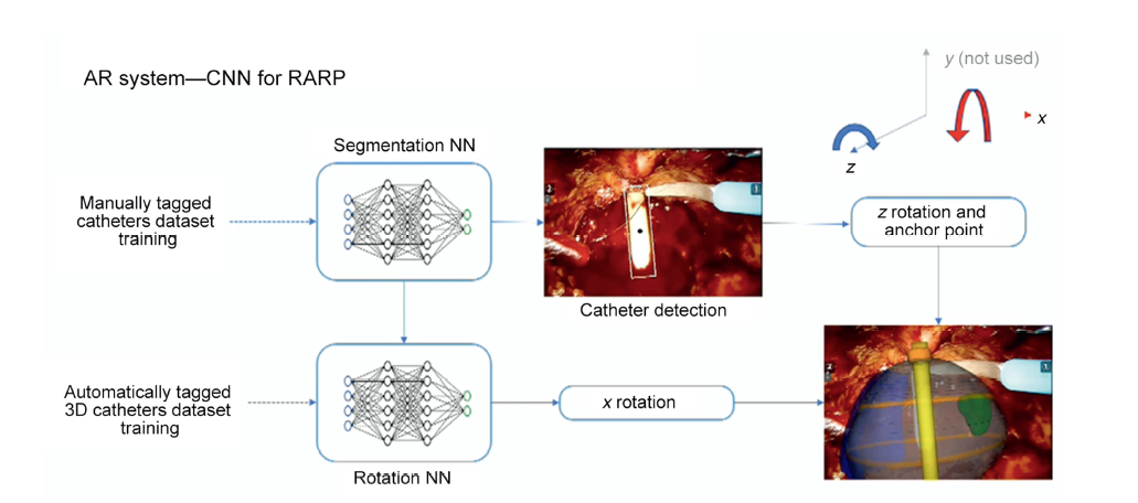
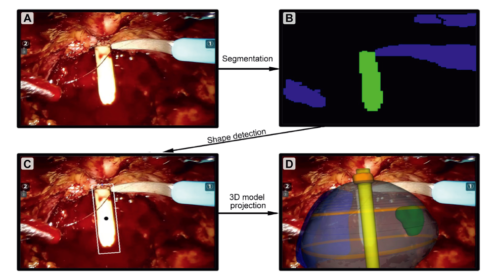
Objective: To evaluate the accuracy of our new three-dimensional (3D) automatic augmented reality (AAR) system guided by artificial intelligence in the identification of tumour's location at the level of the preserved neurovascular bundle (NVB) at the end of the extirpative phase of nerve-sparing robot-assisted radical prostatectomy.
Methods: In this prospective study, we enrolled patients with prostate cancer (clinical stages cT1c-3, cN0, and cM0) with a positive index lesion at target biopsy, suspicious for capsular contact or extracapsular extension at preoperative multiparametric magnetic resonance imaging. Patients underwent robot-assisted radical prostatectomy at San Luigi Gonzaga Hospital (Orbassano, Turin, Italy), from December 2020 to December 2021. At the end of extirpative phase, thanks to our new AAR artificial intelligence driven system, the virtual prostate 3D model allowed to identify the tumour's location at the level of the preserved NVB and to perform a selective excisional biopsy, sparing the remaining portion of the bundle. Perioperative and postoperative data were evaluated, especially focusing on the positive surgical margin (PSM) rates, potency, continence recovery, and biochemical recurrence.
Results: Thirty-four patients were enrolled. In 15 (44.1%) cases, the target lesion was in contact with the prostatic capsule at multiparametric magnetic resonance imaging (Wheeler grade L2) while in 19 (55.9%) cases extracapsular extension was detected (Wheeler grade L3). 3D AAR guided biopsies were negative in all pathological tumour stage 2 (pT2) patients while they revealed the presence of cancer in 14 cases in the pT3 cohort (14/16; 87.5%). PSM rates were 0% and 7.1% in the pathological stages pT2 and pT3 (<3 mm, Gleason score 3), respectively.
Conclusion: With the proposed 3D AAR system, it is possible to correctly identify the lesion's location on the NVB in 87.5% of pT3 patients and perform a 3D-guided tailored nerve-sparing even in locally advanced diseases, without compromising the oncological safety in terms of PSM rates.
. [J]. Asian Journal of Urology, 2023, 10(4): 407-415.
Enrico Checcucci, Alberto Piana, Gabriele Volpi, Pietro Piazzolla, Daniele Amparore, Sabrina De Cillis, Federico Piramide, Cecilia Gatti, Ilaria Stura, Enrico Bollito, Federica Massa, Michele Di Dio, Cristian Fiori, Francesco Porpiglia. Three-dimensional automatic artificial intelligence driven augmented-reality selective biopsy during nerve-sparing robot-assisted radical prostatectomy: A feasibility and accuracy study. Asian Journal of Urology, 2023, 10(4): 407-415.
Checcucci E, Porpiglia F. The future of robotic radical prostatectomy driven by artificial intelligence. Mini-invasive Surg 2021; 5:49. https://doi.org/10.20517/2574-1225.2021.98.
[2]
Sooriakumaran P, Dev HS, Skarecky D, Ahlering T. The importance of surgical margins in prostate cancer. J Surg Oncol 2016; 113:310-5.
doi: 10.1002/jso.24109
pmid: 27004601
[3]
Checcucci E, Amparore D, De Luca S, Autorino R, Fiori C, Porpiglia F. Precision prostate cancer surgery: an overview of new technologies and techniques. Minerva Urol Nefrol 2019; 71:487-501.
doi: 10.23736/S0393-2249.19.03365-4
pmid: 30700084
[4]
Porpiglia F, Checcucci E, Amparore D, Autorino R, Piana A, Bellin A, et al. Augmented-reality robot-assisted radical prostatectomy using hyper-accuracy three-dimensional reconstruction (HA3D_) technology: a radiological and pathological study. BJU Int 2019; 123:834-45.
doi: 10.1111/bju.2019.123.issue-5
[5]
Russo F, Regge D, Armando E, Giannini V, Vignati A, Mazzetti S, et al. Detection of prostate cancer index lesions with multiparametric magnetic resonance imaging (mp-MRI) using whole-mount histological sections as the reference standard. BJU Int 2016; 118:84-94.
doi: 10.1111/bju.13234
pmid: 26198404
[6]
Barentsz JO, Richenberg J, Clements R, Choyke P, Verma S, Villeirs G, et al.; European Society of Urogenital Radiology. ESUR prostate MR guidelines 2012. Eur Radiol 2012; 22:746-57.
doi: 10.1007/s00330-011-2377-y
pmid: 22322308
[7]
Barentsz JO, Weinreb JC, Verma S, Thoeny HC, Tempany CM, Shtern F, et al. Synopsis of the PI-RADS v2 guidelines for multiparametric prostate magnetic resonance imaging and recommendations for use. Eur Urol 2016; 69:41-9.
doi: 10.1016/j.eururo.2015.08.038
pmid: 26361169
[8]
Porpiglia F, Bertolo R, Checcucci E, Amparore D, Autorino R, Dasgupta P, et al.; ESUT Research Group. Development and validation of 3D printed virtual models for robot-assisted radical prostatectomy and partial nephrectomy: urologists’ and patients’ perception. World J Urol 2018; 36:201-7.
doi: 10.1007/s00345-017-2126-1
[9]
Porpiglia F, Bertolo R, Manfredi M, De Luca S, Checcucci E, Morra I, et al. Total anatomical reconstruction during robotassisted radical prostatectomy: implications on early recovery of urinary continence. Eur Urol 2016; 69:485-95.
doi: 10.1016/j.eururo.2015.08.005
[10]
Manfredi M, Checcucci E, Fiori C, Garrou D, Aimar R, Amparore D, et al. Total anatomical reconstruction during robot-assisted radical prostatectomy: focus on urinary continence recovery and related complications after 1000 procedures. BJU Int 2019; 124:477-86.
doi: 10.1111/bju.14716
pmid: 30801887
[11]
Montironi R, Lopez-Beltran A, Mazzucchelli R, Scarpelli M, Bollito E. Assessment of radical prostatectomy specimens and diagnostic reporting of pathological findings. Pathologica 2001; 93:226-32.
pmid: 11433618
[12]
Varma M, Morgan JM. The weight of the prostate gland is an excellent surrogate for gland volume. Histopathology 2010; 57: 55-8.
doi: 10.1111/j.1365-2559.2010.03591.x
pmid: 20653780
[13]
Epstein JI, Egevad L, Amin MB, Delahunt B, Srigley JR, Humphrey PA. The 2014 International Society of Urological Pathology (ISUP) consensus conference on Gleason grading of prostatic carcinoma: definition of grading patterns and proposal for a new grading system. Am J Surg Pathol 2016; 40: 244-52.
doi: 10.1097/PAS.0000000000000530
[14]
Wheeler TM, Dillioglugil O, Kattan MW, Arakawa A, Soh S, Suyama K, et al. Clinical and pathological significance of the level and extent of capsular invasion in clinical stage T1e2 prostate cancer. Hum Pathol 1998; 29:856-62.
doi: 10.1016/s0046-8177(98)90457-9
pmid: 9712429
[15]
Montorsi F, Wilson TG, Rosen RC, Ahlering TE, Artibani W, Carroll PR, et al. Best practices in robot-assisted radical prostatectomy: recommendations of the Pasadena Consensus Panel. Eur Urol 2012; 62:368-81.
doi: 10.1016/j.eururo.2012.05.057
pmid: 22763081
[16]
Clavien PA, Barkun J, de Oliveira ML, Vauthey JN, Dindo D, Schulick RD, et al. The Clavien-Dindo classification of surgical complications: five-year experience. Ann Surg 2009; 250: 187-96.
doi: 10.1097/SLA.0b013e3181b13ca2
pmid: 19638912
[17]
Rosen RC, Riley A, Wagner G, Osterloh IH, Kirkpatrick J, Mishra A. The International Index of Erectile Function (IIEF): a multidimensional scale for assessment of erectile dysfunction. Urology 1997; 49:822-30.
doi: 10.1016/s0090-4295(97)00238-0
pmid: 9187685
[18]
Porpiglia F, Checcucci E, Amparore D, Piazzolla P, Manfredi M, Pecoraro A, et al. Artificial intelligence guided 3D automatic augmented-reality images allow to identify the extracapsular extension on neurovascular bundles during robotic prostatectomy. Eur Urol 2021; 79:S1560. https://doi.org/10.1016/S0302-2838(21)01471-8.
doi: 10.1016/S0302-2838(21)01471-8
[19]
Martini A, Gandaglia G, Fossati N, Scuderi S, Bravi CA, Mazzone E, et al. Defining clinically meaningful positive surgical margins in patients undergoing radical prostatectomy for localised prostate cancer. Eur Urol Oncol 2021; 4:42-8.
doi: 10.1016/j.euo.2019.03.006
pmid: 31411971
[20]
Dinneen E, Haider A, Grierson J, Freeman A, Oxley J, Briggs T, et al. NeuroSAFE frozen section during robot-assisted radical prostatectomy (RARP): peri-operative and histopathological outcomes from the NeuroSAFE PROOF feasibility randomised controlled trial. BJU Int 2021; 127:676-86.
doi: 10.1111/bju.v127.6
[21]
Rocco B, Sighinolfi MC, Cimadamore A, Bonetti LR, Bertoni L, Puliatti S, et al. Digital frozen section of the prostate surface during radical prostatectomy: a novel approach to evaluate surgical margins. BJU Int 2020; 126:336-8.
doi: 10.1111/bju.15108
pmid: 32401370
[22]
Lee S, Kim KB, Jo JK, Ho JN, Oh JJ, Jeong SJ, et al. Prognostic value of focal positive surgical margins after radical prostatectomy. Clin Genitourin Cancer 2016; 14:e313-9. https://doi.org/10.1016/j.clgc.2015.12.013.
doi: 10.1016/j.clgc.2015.12.013
[23]
Checcucci E, Pecoraro A, Amparore D, De Cillis S, Granato S, Volpi G, et al.; Uro-technology and SoMe Working Group of the Young Academic Urologists Working Party of the European Association of Urology. The impact of 3D models on positive surgical margins after robot-assisted radical prostatectomy. World J Urol 2022; 40:2221-9.
doi: 10.1007/s00345-022-04038-8
pmid: 35790535
[24]
Pedraza AM, Wagaskar V, Parekh S, Tewari A. Technical advances in nerve-sparing and continence preservation. Curr Opin Urol 2022; 32:204-10.
doi: 10.1097/MOU.0000000000000958
[25]
Kumar A, Patel VR, Panaiyadiyan S, Seetharam Bhat KR, Moschovas MC, Nayak B. Nerve-sparing robot-assisted radical prostatectomy: current perspectives. Asian J Urol 2021; 8:2-13.
doi: 10.1016/j.ajur.2020.05.012
pmid: 33569267
[26]
Checcucci E, Piazza P, Micali S, Ghazi A, Mottrie A, Porpiglia F, et al.; Uro-technology, SoMe Working Group of the Young Academic Urologists of the European Association of Urology. Threedimensional model reconstruction: the need for standardization to drive tailored surgery. Eur Urol 2022; 81:129-31.
doi: 10.1016/j.eururo.2021.11.010
[27]
Porpiglia F, Checcucci E, Amparore D, Manfredi M, Massa F, Piazzolla P, et al. Three-dimensional elastic augmentedreality robot-assisted radical prostatectomy using hyperaccuracy three-dimensional reconstruction technology: a step further in the identification of capsular involvement. Eur Urol 2019; 76:505-14.
doi: 10.1016/j.eururo.2019.03.037
[28]
Roberts S, Desai A, Checcucci E, Puliatti S, Taratkin M, Kowalewski KF, et al. "Augmented reality" applications in urology: a systematic review. Minerva Urol Nephrol 2022; 74: 528-37.
doi: 10.23736/S2724-6051.22.04726-7
pmid: 35383432
[29]
Gmóez Rivas J, Toribio Vázquez C, Ballesteros Ruiz C, Taratkin M, Marenco JL, Cacciamani GE, et al. [Artificial intelligence and simulation in urology]. Actas Urol Esp 2021; 45: 524-9. [Article in English, Spanish].
doi: 10.1016/j.acuro.2020.10.012
[30]
Checcucci E, De Cillis S, Granato S, Chang P, Afyouni AS, Okhunov Z; Uro-technology and SoMe Working Group of the Young Academic Urologists Working Party of the European Association of Urology. Applications of neural networks in urology: a systematic review. Curr Opin Urol 2020; 30: 788-807.
doi: 10.1097/MOU.0000000000000814
[31]
Checcucci E, Autorino R, Cacciamani GE, Amparore D, De Cillis S, Piana A, et al.; Uro-technology and SoMe Working Group of the Young Academic Urologists Working Party of the European Association of Urology. Artificial intelligence and neural networks in urology: current clinical applications. Minerva Urol Nefrol 2020; 72:49-57.
doi: 10.23736/S0393-2249.19.03613-0
pmid: 31833725
[32]
Amparore D, Checcucci E, Piazzolla P, Piramide F, De Cillis S, Piana A, et al. Indocyanine green drives computer vision based 3D augmented reality robot assisted partial nephrectomy: the beginning of "automatic" overlapping era. Urology 2022; 164: e312-6. https://doi.org/10.1016/j.urology.2021.10.053.
doi: 10.1016/j.urology.2021.10.053
[33]
Piana A, Gallioli A, Amparore D, Diana P, Territo A, Campi R, et al. Three-dimensional augmented reality-guided roboticassisted kidney transplantation: breaking the limit of atheromatic plaques. Eur Urol 2022; 82:419-26.
doi: 10.1016/j.eururo.2022.07.003
[34]
Moglia A, Georgiou K, Georgiou E, Satava RM, Cuschieri A. A systematic review on artificial intelligence in robot-assisted surgery. Int J Surg 2021 Nov; 95:106151. https://doi.org/10.1016/j.ijsu.2021.106151.
doi: 10.1016/j.ijsu.2021.106151