|
|
|
| Transforming urinary stone disease management by artificial intelligence-based methods: A comprehensive review |
Anastasios Anastasiadisa,Antonios Koudonasa,Georgios Langasa,Stavros Tsiakarasa,*( ),Dimitrios Memmosa,Ioannis Mykoniatisa,Evangelos N. Symeonidisa,Dimitrios Tsiptsiosb,Eliophotos Savvidesc,Ioannis Vakalopoulosa,Georgios Dimitriadisa,Jean de la Rosetted ),Dimitrios Memmosa,Ioannis Mykoniatisa,Evangelos N. Symeonidisa,Dimitrios Tsiptsiosb,Eliophotos Savvidesc,Ioannis Vakalopoulosa,Georgios Dimitriadisa,Jean de la Rosetted
|
a 1st Department of Urology, Aristotle University of Thessaloniki, School of Medicine, “G.Gennimatas” General Hospital, Thessaloniki, Greece
b Neurology Department, Democritus University of Thrace, Alexandroupolis, Greece
c Department of Urology, Main Kinzig Kliniken, Gelnhausen, Germany
d Department of Urology, Istanbul Medipol Mega University Hospital, Istanbul, Turkey |
|
|
|
|
Abstract Objective To provide a comprehensive review on the existing research and evidence regarding artificial intelligence (AI) applications in the assessment and management of urinary stone disease.
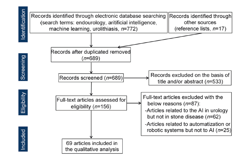
Methods A comprehensive literature review was performed using PubMed, Scopus, and Google Scholar databases to identify publications about innovative concepts or supporting applications of AI in the improvement of every medical procedure relating to stone disease. The terms ‘‘endourology’’, ‘‘artificial intelligence’’, ‘‘machine learning’’, and ‘‘urolithiasis'’ were used for searching eligible reports, while review articles, articles referring to automated procedures without AI application, and editorial comments were excluded from the final set of publications. The search was conducted from January 2000 to September 2023 and included manuscripts in the English language.
Results A total of 69 studies were identified. The main subjects were related to the detection of urinary stones, the prediction of the outcome of conservative or operative management, the optimization of operative procedures, and the elucidation of the relation of urinary stone chemistry with various factors.
Conclusion AI represents a useful tool that provides urologists with numerous amenities, which explains the fact that it has gained ground in the pursuit of stone disease management perfection. The effectiveness of diagnosis and therapy can be increased by using it as an alternative or adjunct to the already existing data. However, little is known concerning the potential of this vast field. Electronic patient records, containing big data, offer AI the opportunity to develop and analyze more precise and efficient diagnostic and treatment algorithms. Nevertheless, the existing applications are not generalizable in real-life practice, and high-quality studies are needed to establish the integration of AI in the management of urinary stone disease.
|
|
Received: 12 September 2022
Available online:
|
|
Corresponding Authors:
Stavros Tsiakaras
E-mail: drstavros90@gmail.com
|
|
|
|
|
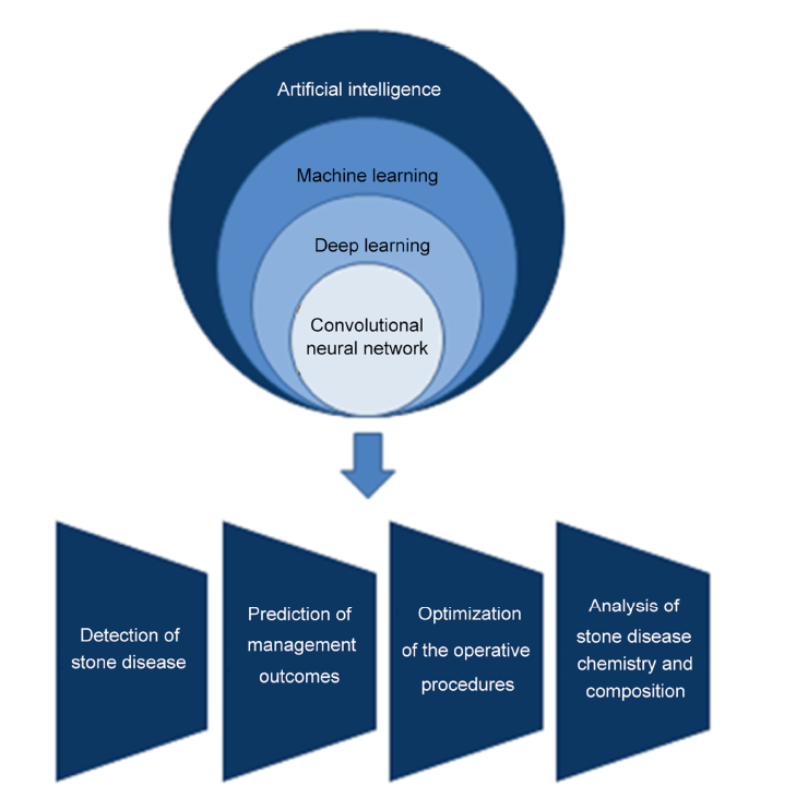
Subsets of artificial intelligence with emergent role in stone disease management.
|
|
|
Flowchart of the literature selection process for articles.
|
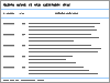
| Study | Objective | Study design | AI-based outcome | Comparator arm outcome | | Li et al. [13] | Detection of urinary stones by CT | Cross-sectional | Detection accuracy of 99.95% | Other algorithms with lower performance | | Parakh et al. [14] | Detection of urinary stones by CT | Cross-sectional | High accuracy in stone detection (AUC of 0.954) | Other algorithms with lower performance | | L?ngkvist et al. [15] | Detection of urinary stones by CT | Cross-sectional | Optimized accuracy with an AUC of 0.9971 | No comparator | | Caglayan et al. [16] | Detection of urinary stones by CT | Cross-sectional | Accuracy of 63%-93%, depending on imaging plane and stone size class | No comparator | | Jendeberg et al. [17] | Differentiation of ureteral stones and pelvic phleboliths by CT | Cross-sectional | Accuracy of 92% | Mean radiologist accuracy: 86%; majority vote accuracy: 93% | | De Perrot et al. [18] | Differentiation of ureteral stones and pelvic phleboliths by CT | Cross-sectional | Overall accuracy of 85.1% (AUC of 0.902) | Other algorithms with lower performance | | Chak et al. [19] | Detection of urinary stones by CT | Cross-sectional | Accuracy of 95%-99%, depending on the number of features used by the algorithm | No comparator | | G P et al. [20] | Detection of urinary stones by CT | Cross-sectional | Accuracy of 96.82% | No comparator | | Elton et al. [21] | Detection of urinary stones by CT | Cross-sectional | High accuracy in stone detection (AUC of 0.95) | No comparator | | Krishna et al. [22] | Differentiation of renal stones and renal cysts by US | Cross-sectional | Accuracy of 98.1% | No comparator | | Balamurugan and Arumugam [23] | Differentiation of renal stones among other abnormalities in US | Cross-sectional | Accuracy of 95.83% | Other algorithms with lower performance | | Selvarani and Rajendran [24] | Detection of renal stones by US | Cross-sectional | Accuracy of 98.8% | Other algorithms with lower performance | | Viswanath et al. [25] | Detection of renal stones by US | Cross-sectional | Accuracy of 98.9% | Other algorithms with lower performance | | Akkasaligar and Biradar [26] | Detection of renal stones by US | Cross-sectional | Accuracy of 96.8% | No comparator | | Verma et al. [27] | Detection of renal stones by US | Cross-sectional | Accuracy of 89% | Other algorithms with lower performance | | Kobayashi et al. [28] | Detection of radio-opaque urinary stones in KUB X-ray images | Cross-sectional | Sensitivity and PPV of 89.6% and 56.9% for the kidney, 92.5% and 87.6% for the proximal ureter, 59.1% and 50% for the mid-ureter, 80% and 55.8% for the distal ureter | No comparator | | Aksakalli et al. [29] | Detection of radio-opaque urinary stones in KUB X-ray images | Cross-sectional | Precision of 78.4% | Other algorithms with lower performance |
|
|
Summary of studies regarding AI in the detection of stone disease.
|
| Study | Objective | Study design | AI-based outcome | Comparator arm outcome | | Cummings et al. [30] | Prediction of SSP | Case-control | Accuracy of 76% | No comparator | | Dal Moro et al. [31] | Prediction of SSP | Case-control | 84.5% sensitivity and 86.9% specificity | Other algorithms with lower performance | | Solakhan et al. [32] | Prediction of SSP | Case-control | Accuracy of 92.8% | Other algorithms with lower performance | | Park et al. [33] | Prediction of SSP | Case-control | AUCs of 0.859 (stones of <5 mm) and 0.881 (stones of 5-10 mm) | AUC of 0.847 (stones of <5 mm) and 0.817 (stones of 5 mm-10 mm) | | Poulakis et al. [34] | Prediction of lower pole clearance after ESWL | Case-control | Accuracy of 92% | No comparator | | Gomha et al. [35] | Prediction of clearance after ESWL for ureteral stones | Case-control | Accuracy of 77.7% | Accuracy of 93.2% | | Moorthy and Krishnan [36] | Prediction of renal stone fragmentation after ESWL | Case-control | Accuracy of 90% | No comparator | | Choo et al. [37] | Prediction of clearance after ESWL for ureteral stones | Case-control | Accuracy of 92.29% | No comparator | | Seckiner et al. [38] | Prediction of clearance after ESWL for renal stones | Case-control | Accuracy of 88.70% | No comparator | | Mannil et al. [39] | Prediction of renal stones fragmentation after ESWL | Case-control | AUC of 0.85 | Other algorithms with lower performance | | Yang et al. [40] | Prediction of clearance after ESWL for renal or upper ureter stones | Case-control | AUC of 0.85 for stone-free status in an interval of 4 weeks; AUC of 0.78 for stone-free status after single session ESWL | Other algorithms with similar performance | | Tsitsiflis et al. [41] | Prediction of complications after ESWL for renal or ureteral stones | Case-control | Accuracy of 81.43% | No comparator | | Handa et al. [42] | Quantification of ESWL-induced renal injury by MRI | Experimental | Strong correlation between model prediction and morphology (r=0.9691) | No comparator | | Aminsharifi et al. [43] | Prediction of multiple outcomes after PCNL | Case-control | Accuracy of 91.8%, 83% regarding stone clearance and need for blood transfusion; AUC of 0.915 for stone clearance | AUCs of 0.615 and 0.621 for stone clearance according to GSS and CROES nomograms | | Shabaniyan et al. [44] | Prediction of multiple outcomes after PCNL | Case-control | Accuracy of 94.8% in prediction of the procedures‘ outcome, 85.2% accuracy in predicting the need for stent placement and 95% in predicting blood transfusion | Multiple decision support systems achieving higher performances in different parameters | | Aminsharifi et al. [45] | Prediction of multiple outcomes after PCNL | Case-control | Accuracy of 82.8%, 92.5%-98.2%, 81.1%, and 85.8% for stone clearance, need for a second procedure, stent insertion by urine extravasation, and blood transfusion | No comparator | | Geraghty et al. [46] | Prediction of multiple outcomes after PCNL | Case-control | Multiple classification models tested, highest accuracy of 99% and AUCs of 0.99-1.00 achieved for need for transfusion and infectious complications | No comparator | | Zhao et al. [47] | Prediction of stone clearance after PCNL | Case-control | AUC of 0.879 | AUC of 0.800 for GSS; AUC of 0.844 for S.T.O.N.E. score | | Chen et al. [48] | Prediction of sepsis after fURS or PCNL for proximal ureteral stones | Case-control | AUC of 0.874 for DNN model | AUC of 0.783 for LASSO model |
|
|
Summary of studies regarding AI in the prediction of management outcomes.
|
| Study | Objective | Study design | AI-based outcome | Comparator arm outcome | | Hamid et al. [49] | Optimization of ESWL protocol | Cross-sectional | Accuracy of 75% for predicting shockwave number and 100% for predicting patients needing shockwave number beyond protocol | No comparator | | Goyal et al. [50] | Optimization of ESWL protocol | Cross-sectional | Correlation coefficients for power level and shockwave number of 0.8343 and 0.9329, respectively | Correlation coefficients for power level and shockwave number 0.0195 and 0.5726, respectively | | Mannil et al. [51] | Optimization of ESWL protocol | Experimental | AUC of 0.838 in the prediction of fragmentation with less than 72 shockwaves | Other multivariate models with lower performance | | Chen et al. [52] | Optimization of ESWL protocol | Cross-sectional | Prediction accuracy values for power level, shockwave rate of 98.8%, 98.1%, respectively | Other multivariate models with lower performance | | Muller et al. [53] | Optimization of ESWL protocol | Cross-sectional | Shockwave hit rate of 75.3% | Shockwave hit rate of 55.2% | | Taguchi et al. [54] | Optimization of PCNL puncture | Experimental | Puncture success rate of 100%; puncture time of 35 s | Puncture success rate of 70.6%; puncture time of 46 s | | Wang et al. [55] | Optimization of PCNL puncture | Experimental | Average recognition precision of 79% (SE: 4%) for cortex, 85% (SE: 6%) for medulla, and 91% (SE: 5%) for calyx | No comparator | | Li et al. [56] | Optimization of PCNL puncture | Cross-sectional | ANN model achieved a better localization and puncture method selection compared to the MVRA model and the surgeon's experience | No comparator | | Jeong et al. [57] | Optimization of RIRS safety profile | Experimental | Recognition of tissue exposure to laser energy with accuracy of 95% and latency time of 0.5 s | No comparator |
|
|
Summary of studies regarding the contribution of AI in the optimization of the operative procedure.
|
| Study | Objective | Study design | AI-based outcome | Comparator arm outcome | | Dussol et al. [58] | Risk factors for calcium stones | Case-control | Classification accuracy between stone formers and controls: 74.4% | 75.8% | | Dussol et al. [59] | Risk factors for calcium stones | Case-control | CaOx supersaturation and 24 h-urea for all men and women with a family history | No comparator | | Kazemi and Mirroshandel [60] | Risk of nephrolithiasis | Cohort | Accuracy of 97.1% | Other classifiers with lower accuracy | | Chen et al. [61] | Risk of forming renal stones of >2 cm | Cohort | AUC of 0.69 | AUC of 0.74 | | Kavoussi et al. [62] | Prediction of 24 h urine abnormalities relevant for stone disease | Cohort | Higher accuracy in prediction of urine volume, uric acid, and natrium abnormalities | Higher accuracy in prediction of pH and citrate abnormalities | | Caudarella et al. [63] | Risk of stone disease recurrence | Case-control | Accuracy of 88.8% | Accuracy of 67.5% | | Chiang et al. [64] | Risk for stone disease | Case-control | Accuracy of 89% | Accuracy of 74% | | Xiang et al. [65] | Identification of CaOx crystallization in urine sediment | Cross-sectional | Accuracy of 74% | Accuracy of 74% | | Kletzmayr et al. [66] | Recognition of crystallization inhibition | Experimental | IP6 analogues inhibit effectively CaOx crystallization | No comparator | | Kriegshauser et al. [67] | Stone composition by CT | Cross-sectional | Accuracy of 97% (UA instead of non-UA stones) and 72% among non-UA stones | Other multivariate models with lower performance | | Kriegshauser et al. [68] | Stone composition by CT | Cross-sectional | Accuracy of 100% (UA instead of non-UA stones) and 88% among non-UA stones | Other multivariate models with lower performance | | Zhang et al. [69] | Stone composition by CT | Cross-sectional | AUC of 0.965 (SD: 0.029) for UA instead of non-UA stones | Sensitivity of 94.4% and specificity of 93.7% for model using CT TA | | Gro?e Hokamp et al. [70] | Stone composition by CT | Cross-sectional | Accuracy of 91.1% on a per-voxel basis; accuracy of 87.1%-90.4% on independently tested acquisitions | No comparator | | Tang et al. [71] | Stone composition by CT | Cross-sectional | Accuracy of 88.3% for COM instead of non-COM stones (AUC=0.933) | No comparator | | Black et al. [72] | Stone composition by visual image | Cross-sectional | Prediction precision for each stone composition from 71.43% (struvite) to 95% (COM stones) | No comparator | | Lopez et al. [73] | Stone composition by visual image | Cross-sectional | Precision of 93%-98%, depending on stone type | Other multivariate models with lower performance | | El Beze et al. [74] | Stone composition by visual image | Cross-sectional | PPV of 96%-99%, depending on stone type | PPV of 88%-99%, depending on stone type | | Ochoa-Ruiz et al. [75] | Stone composition by visual image | Cross-sectional | Overall precision of 97% | Overall precision of 96% | | Mendez-Ruiz et al. [76] | Stone composition by visual image | Cross-sectional | Overall accuracy of 74.38% and 88.52%, depending on the image capturing method | Overall accuracy of 45% | | Kim et al. [77] | Stone composition by visual image | Cross-sectional | AUC of 0.98-1.00, depending on stone type | Other multivariate models with lower performance | | Fitri et al. [78] | Stone composition by microtomography | Cross-sectional | Overall accuracy of 99.59% | No comparator | | Sa?l? et al. [79] | Stone composition by dielectric properties | Cross-sectional | Overall accuracy of 98.17% | No comparator | | Cui et al. [80] | Stone composition by Raman spectroscopy | Cross-sectional | Overall accuracy of 96.3% | No comparator | | Onal and Tekgul [81] | Stone composition by smartphone microscopy | Cross-sectional | Overall accuracy of 88% | No comparator |
|
|
Summary of studies on the contribution of AI in the elucidation of stone disease chemistry and composition.
|
| [1] |
Malik P, Pathania M, Rathaur VK. Overview of artificial intelligence in medicine. J Fam Med Prim Care 2019; 8:2328-31.
|
| [2] |
Mintz Y, Brodie R. Introduction to artificial intelligence in medicine. Minim Invasive Ther Allied Technol 2019; 28:73-81.
doi: 10.1080/13645706.2019.1575882
|
| [3] |
Kueper JK. Primer for artificial intelligence in primary care. Can Fam Physician 2021; 67:889-93.
doi: 10.46747/cfp.6712889
pmid: 34906934
|
| [4] |
Schmidhuber J. Deep learning in neural networks: an overview. Neural Network 2015; 61:85-117.
doi: 10.1016/j.neunet.2014.09.003
|
| [5] |
Frankish K, Ramsey WM. The Cambridge handbook of artificial intelligence. Cambridge: Cambridge University Press; 2014. p. 151-66.
|
| [6] |
Rowe M. An introduction to machine learning for clinicians. Acad Med 2019; 94:1433-6.
doi: 10.1097/ACM.0000000000002792
pmid: 31094727
|
| [7] |
Choi RY, Coyner AS, Kalpathy-Cramer J, Chiang MF, Campbell JP. Introduction to machine learning, neural networks, and deep learning. Transl Vis Sci Technol 2020; 9:14. https://doi.org/10.1167/tvst.9.2.14.
|
| [8] |
Rabhi S, Jakubowicz J, Metzger MH. Deep learning versus conventional machine learning for detection of healthcareassociated infections in French clinical narratives. Methods Inf Med 2019; 58:31-41.
doi: 10.1055/s-0039-1677692
|
| [9] |
Yamashita R, Nishio M, Do RKG, Togashi K. Convolutional neural networks: an overview and application in radiology. Insights Imaging 2018; 9:611-29.
doi: 10.1007/s13244-018-0639-9
|
| [10] |
Jin KH, McCann MT, Froustey E, Unser M. Deep convolutional neural network for inverse problems in imaging. IEEE Trans Image Process 2017; 26:4509-22.
doi: 10.1109/TIP.2017.2713099
|
| [11] |
Huang JA, Hartanti IR, Colin MN, Pitaloka DA. Telemedicine and artificial intelligence to support self-isolation of COVID-19 patients: recent updates and challenges. Digital Health 2022; 8. 20552076221100634. https://doi.org/10.1177/20552076221100634.
|
| [12] |
Hamet P, Tremblay J. Artificial intelligence in medicine. Metabolism 2017; 69:S36-40. https://doi.org/10.1016/j.metabol.2017.01.011.
doi: 10.1016/j.metabol.2017.01.011
|
| [13] |
Li D, Xiao C, Liu Y, Chen Z, Hassan H, Su L, et al. Deep Segmentation Networks for segmenting kidneys and detecting kidney stones in unenhanced abdominal CT images. Diagnostics 2022; 12:1788. https://doi.org/10.3390/diagnostics12081788.
doi: 10.3390/diagnostics12081788
|
| [14] |
Parakh A, Lee H, Lee JH, Eisner BH, Sahani DV, Do S. Urinary stone detection on CT images using deep convolutional neural networks: evaluation of model performance and generalization. Radiol Artif Intell 2019; 1:e180066. https://doi.org/10.1148/ryai.2019180066.
|
| [15] |
L?ngkvist M, Jendeberg J, Thunberg P, Loutfi A, Lidén M. Computer aided detection of ureteral stones in thin slice computed tomography volumes using Convolutional Neural Networks. Comput Biol Med 2018;97:153-60.
|
| [16] |
Caglayan A, Horsanali MO, Kocadurdu K, Ismailoglu E, Guneyli S. Deep learning model-assisted detection of kidney stones on computed tomography. Int Braz J Urol 2022; 48: 830-9.
doi: 10.1590/S1677-5538.IBJU.2022.0132
pmid: 35838509
|
| [17] |
Jendeberg J, Thunberg P, Lidén M. Differentiation of distal ureteral stones and pelvic phleboliths using a convolutional neural network. Urolithiasis 2021; 49:41-9.
doi: 10.1007/s00240-020-01180-z
|
| [18] |
De Perrot T, Hofmeister J, Burgermeister S, Martin SP, Feutry G, Klein J, et al. Differentiating kidney stones from phleboliths in unenhanced low-dose computed tomography using radiomics and machine learning. Eur Radiol 2019; 29:4776-82.
doi: 10.1007/s00330-019-6004-7
pmid: 30747299
|
| [19] |
Chak P, Navadiya P, Parikh B, Pathak KC. Neural network and svm based kidney stone based medical image classification. In: International conference on computer vision and image processing. Singapore: Springer; 2019. p158-73.
|
| [20] |
G P VP, Reddy KVS, Kiruthik AM, ArunNehru J. Prediction of kidney stones using machine learning. Int J Res Appl Sci Eng Technol 2022; 10:1037-44.
|
| [21] |
Elton DC, Turkbey EB, Pickhardt PJ, Summers RM.A deep learning system for automated kidney stone detection and volumetric segmentation on noncontrast CT scans. Med Phys 2022; 49:2545-54.
|
| [22] |
Krishna KD, Akkala V, Bharath R, Rajalakshmi P, Mohammed A, Merchant S, et al. Computer aided abnormality detection for kidney on FPGA based IoT enabled portable ultrasound imaging system. IRBM 2016; 37:189-97.
doi: 10.1016/j.irbm.2016.05.001
|
| [23] |
Balamurugan SP, Arumugam G. A novel method for predicting kidney diseases using optimal artificial neural network in ultrasound images. IJIE 2020; 7:37-55.
|
| [24] |
Selvarani S, Rajendran P. Detection of renal calculi in ultrasound image using meta-heuristic support vector machine. J Med Syst 2019; 43:1-9.
doi: 10.1007/s10916-018-1115-2
|
| [25] |
Viswanath K, Anilkumar B, Gunasundari R. Design of deep learning reactionediffusion level set segmentation approach for health care related to automatic kidney stone detection analysis. Multimed Tool Appl 2022:1-43.
|
| [26] |
Akkasaligar PT, Biradar S.Diagnosis of renal calculus disease in medical ultrasound images. In: 2016 IEEE international conference on computational intelligence and computing research (ICCIC). IEEE; 2016. p. 1-5. https://doi.org/10.1109/ICCIC.2016.7919642.
|
| [27] |
Verma J, Nath M, Tripathi P, Saini K. Analysis and identification of kidney stone using kth nearest neighbour (KNN) and support vector machine (SVM) classification techniques. Pattern Recogn Image Anal 2017; 27:574-80.
doi: 10.1134/S1054661817030294
|
| [28] |
Kobayashi M, Ishioka J, Matsuoka Y, Fukuda Y, Kohno Y, Kawano K, et al. Computer-aided diagnosis with a convolutional neural network algorithm for automated detection of urinary tract stones on plain X-ray. BMC Urol 2021; 21:1-10.
doi: 10.1186/s12894-020-00770-8
|
| [29] |
Aksakalli I, Ka?dio?lu S, Hanay YS. Kidney X-ray images classification using machine learning and deep learning methods. Balkan Journal of Electrical and Computer Engineering 2021;9: 144-51.
|
| [30] |
Cummings JM, Boullier JA, Izenberg SD, Kitchens DM, Kothandapani RV. Prediction of spontaneous ureteral calculous passage by an artificial neural network. J Urol 2000; 164:326-8.
pmid: 10893576
|
| [31] |
Dal Moro F, Abate A, Lanckriet G, Arandjelovic G, Gasparella P, Bassi P, et al. A novel approach for accurate prediction of spontaneous passage of ureteral stones: support vector machines. Kidney Int 2006; 69:157-60.
pmid: 16374437
|
| [32] |
Solakhan M, Seckiner SU, Seckiner I. A neural network-based algorithm for predicting the spontaneous passage of ureteral stones. Urolithiasis 2020; 48:527-32.
doi: 10.1007/s00240-019-01167-5
|
| [33] |
Park JS, Kim DW, Lee D, Lee T, Koo KC, Han WK, et al. Development of prediction models of spontaneous ureteral stone passage through machine learning: comparison with conventional statistical analysis. PLoS One 2021; 16:e0260517. https://doi.org/10.1371/JOURNAL.PONE.0260517.
|
| [34] |
Poulakis V, Dahm P, Witzsch U, De Vries R, Remplik J, Becht E. Prediction of lower pole stone clearance after shock wave lithotripsy using an artificial neural network. J Urol 2003; 169: 1250-6.
doi: 10.1097/01.ju.0000055624.65386.b9
pmid: 12629337
|
| [35] |
Gomha MA, Sheir KZ, Showky S, Abdel-Khalek M, Mokhtar AA, Madbouly K. Can we improve the prediction of stone-free status after extracorporeal shock wave lithotripsy for ureteral stones? A neural network or a statistical model? J Urol 2004; 172:175-9.
|
| [36] |
Moorthy K, Krishnan M. Prediction of fragmentation of kidney stones: a statistical approach from NCCTimages. Can Urol Assoc J 2016; 10:E237-40. https://doi.org/10.5489/cuaj.3674.
|
| [37] |
Choo MS, Uhmn S, Kim JK, Han JH, Kim D-H, Kim J, et al. A prediction model using machine learning algorithm for assessing stone-free status after single session shock wave lithotripsy to treat ureteral stones. J Urol 2018; 200:1371-7.
doi: S0022-5347(18)43554-9
pmid: 30036513
|
| [38] |
Seckiner I, Seckiner S, Sen H, Bayrak O, Dogan K, Erturhan S. A neural network-based algorithm for predicting stone-free status after ESWL therapy. Int Braz J Urol 2017; 43:1110-4.
doi: 10.1590/S1677-5538.IBJU.2016.0630
pmid: 28727384
|
| [39] |
Mannil M, von Spiczak J, Hermanns T, Poyet C, Alkadhi H, Fankhauser CD. Three-dimensional texture analysis with machine learning provides incremental predictive information for successful shock wave lithotripsy in patients with kidney stones. J Urol 2018; 200:829-36.
doi: S0022-5347(18)42986-2
pmid: 29673945
|
| [40] |
Yang SW, Hyon YK, Na HS, Jin L, Lee JG, Park JM, et al. Machine learning prediction of stone-free success in patients with urinary stone after treatment of shock wave lithotripsy. BMC Urol 2020; 20:1-8.
doi: 10.1186/s12894-019-0555-4
|
| [41] |
Tsitsiflis A, Kiouvrekis Y, Chasiotis G, Perifanos G, Gravas S, Stefanidis I, et al. The use of an artificial neural network in the evaluation of the extracorporeal shockwave lithotripsy as a treatment of choice for urinary lithiasis. Asian J Urol 2022; 9: 132-8.
doi: 10.1016/j.ajur.2021.09.005
pmid: 35509481
|
| [42] |
Handa RK, Territo PR, Blomgren PM, Persohn SA, Lin C, Johnson CD, et al. Development of a novel magnetic resonance imaging acquisition and analysis workflow for the quantification of shock wave lithotripsy-induced renal hemorrhagic injury. Urolithiasis 2017; 45:507-13.
doi: 10.1007/s00240-016-0959-5
pmid: 28074231
|
| [43] |
Aminsharifi A, Irani D, Pooyesh S, Parvin H, Dehghani S, Yousofi K, et al. Artificial neural network system to predict the postoperative outcome of percutaneous nephrolithotomy. J Endourol 2017; 31:461-7.
doi: 10.1089/end.2016.0791
pmid: 28287830
|
| [44] |
Shabaniyan T, Parsaei H, Aminsharifi A, Movahedi MM, Jahromi AT, Pouyesh S, et al. An artificial intelligence-based clinical decision support system for large kidney stone treatment. Australas Phys Eng Sci Med 2019; 42:771-9.
doi: 10.1007/s13246-019-00780-3
|
| [45] |
Aminsharifi A, Irani D, Tayebi S, Jafari Kafash T, Shabanian T, Parsaei H. Predicting the postoperative outcome of percutaneous nephrolithotomy with machine learning system: software validation and comparative analysis with Guy’s Stone Score and the CROES Nomogram. J Endourol 2020; 34:692-9.
doi: 10.1089/end.2019.0475
pmid: 31886708
|
| [46] |
Geraghty R, Finch W, Fowler S, Sriprasad S, Smith D, Dickinson A, et al. Use of internally validated machine and deep learning models to predict outcomes of percutaneous nephrolithotomy using data from the BAUS PCNL audit. medRxiv 2022. https://doi.org/10.1101/2022.06.16.22276481.
|
| [47] |
Zhao H, Li W, Li J, Li L, Wang H, Guo J. Predicting the stonefree status of percutaneous nephrolithotomy with the machine learning system: comparative analysis with Guy’s stone score and the STONE score system. Front Mol Biosci 2022; 9: 880291. https://doi.org/10.3389/fmolb.2022.880291.
doi: 10.3389/fmolb.2022.880291
|
| [48] |
Chen M, Yang J, Lu J, Zhou Z, Huang K, Zhang S, et al. Ureteral calculi lithotripsy for single ureteral calculi: can DNN-assisted model help preoperatively predict risk factors for sepsis? Eur Radiol 2022; 32:8540-9.
|
| [49] |
Hamid A, Dwivedi U, Singh T, Gopi Kishore M, Mahmood M, Singh H, et al. Artificial neural networks in predicting optimum renal stone fragmentation by extracorporeal shock wave lithotripsy: a preliminary study. BJU Int 2003; 91:821-4.
pmid: 12780841
|
| [50] |
Goyal NK, Kumar A, Trivedi S, Dwivedi US, Singh T, Singh PB.A comparative study of artificial neural network and multivariate regression analysis to analyze optimum renal stone fragmentation by extracorporeal shock wave lithotripsy. Saudi J Kidney Dis Transpl 2010; 21:1073-80.
|
| [51] |
Mannil M, Von Spiczak J, Hermanns T, Alkadhi H, Fankhauser CD. Prediction of successful shock wave lithotripsy with CT: a phantom study using texture analysis. Abdom Radiol (NY) 2018; 43:1432-8.
doi: 10.1007/s00261-017-1309-y
pmid: 28840294
|
| [52] |
Chen Z, Zeng DD, Seltzer RG, Hamilton BD. Automated generation of personalized shock wave lithotripsy protocols: treatment planning using deep learning. JMIR Med Inform 2021; 9:e24721. https://doi.org/10.2196/24721.
doi: 10.2196/24721
|
| [53] |
Muller S, Abildsnes H, ?stvik A, Kragset O, Gang?s I, Birke H, et al. Can a dinosaur think? Implementation of artificial intelligence in extracorporeal shock wave lithotripsy. Eur Urol Open Sci 2021;27:33-42.
|
| [54] |
Taguchi K, Hamamoto S, Okada A, Tanaka Y, Sugino T, Unno R, et al. Robot-assisted fluoroscopy versus ultrasound-guided renal access for nephrolithotomy: a phantom model benchtop study. J Endourol 2019; 33:987-94.
doi: 10.1089/end.2019.0432
pmid: 31642340
|
| [55] |
Wang C, Calle P, Ton NBT, Zhang Z, Yan F, Donaldson AM, et al. Deep-learning-aided forward optical coherence tomography endoscope for percutaneous nephrostomy guidance. Biomed Opt Express 2021; 12:2404-18.
doi: 10.1364/BOE.421299
pmid: 33996237
|
| [56] |
Li G, Liu Z, Zhang Y, Chen Z, Hu P, Chen Q, et al. Discrimination analysis of B-mode ultrasonography and X-ray on the percutaneous nephrolithotomy localization of urinary stones: a prospective, controlled study. Int J Clin Exp Med 2016; 9: 2261-8.
|
| [57] |
Jeong J, Chang K, Lee J, Choi J. A warning system for urolithiasis via retrograde intrarenal surgery using machine learning: an experimental study. BMC Urol 2022; 22:1-8.
doi: 10.1186/s12894-021-00951-z
|
| [58] |
Dussol B, Verdier JM, Le Goff JM, Berthezene P, Berland Y. Artificial neural networks for assessing the risk of urinary calcium stone among men. Urol Res 2006; 34:17-25.
|
| [59] |
Dussol B, Verdier JM, Le Goff JM, Berthezene P, Berland Y. Artificial neural networks for assessing the risk factors for urinary calcium stones according to gender and family history of stone. Scand J Urol Nephrol 2007; 41:414-8.
pmid: 17853052
|
| [60] |
Kazemi Y, Mirroshandel SA. A novel method for predicting kidney stone type using ensemble learning. Artif Intell Med 2018; 84:117-26.
doi: S0933-3657(17)30271-3
pmid: 29241659
|
| [61] |
Chen Z, Prosperi M, Bird VG, Bird VY. Analysis of factors associated with large kidney stones: stone composition, comorbid conditions, and 24-h urine parametersda machine learning-aided approach. SN Compr Clin Med 2019; 1: 597-602.
doi: 10.1007/s42399-019-00087-0
|
| [62] |
Kavoussi NL, Floyd C, Abraham A, Sui W, Bejan C, Capra JA, et al. Machine learning models to predict 24 hour urinary abnormalities for kidney stone disease. Urology 2022; 169:52-7.
doi: 10.1016/j.urology.2022.07.008
|
| [63] |
Caudarella R, Tonello L, Rizzoli E, Vescini F. Predicting fiveyear recurrence rates of kidney stones: an artificial neural network model. Arch Ital Urol Androl 2011; 83:14-9.
|
| [64] |
Chiang D, Chiang HC, Chen WC, Tsai FJ. Prediction of stone disease by discriminant analysis and artificial neural networks in genetic polymorphisms: a new method. BJU Int 2003; 91: 661-6.
pmid: 12699480
|
| [65] |
Xiang H, Chen Q, Wu Y, Xu D, Qi S, Mei J, et al. Urine calcium oxalate crystallization recognition method based on deep learning. In: 2019 international conference on automationcomputational and technology management,ICACTM 2019; 2019. p. 30-3. https://doi.org/10.1109/ICACTM.2019.8776769.
|
| [66] |
Kletzmayr A, Mulay SR, Motrapu M, Luo Z, Anders HJ, Ivarsson ME, et al. Inhibitors of calcium oxalate crystallization for the treatment of oxalate nephropathies. Adv Sci 2020; 7: 1903337. https://doi.org/10.1002/advs.201903337.
doi: 10.1002/advs.v7.8
|
| [67] |
Kriegshauser JS, Silva AC, Paden RG, He M, Humphreys MR, Zell SI, et al. Ex vivo renal stone characterization with singlesource dual-energy computed tomography: a multiparametric approach. Acad Radiol 2016; 23:969-76.
doi: 10.1016/j.acra.2016.03.009
pmid: 27212607
|
| [68] |
Kriegshauser JS, Paden RG, He M, Humphreys MR, Zell SI, Fu Y, et al. Rapid kV-switching single-source dual-energy CT ex vivo renal calculi characterization using a multiparametric approach: refining parameters on an expanded dataset. Abdom Radiol (NY) 2018; 43:1439-45.
doi: 10.1007/s00261-017-1331-0
pmid: 28952007
|
| [69] |
Zhang GMY, Sun H, Shi B, Xu M, Xue HD, Jin ZY. Uric acid versus non-uric acid urinary stones: differentiation with single energy CT texture analysis. Clin Radiol 2018; 73:792-9.
doi: 10.1016/j.crad.2018.04.010
|
| [70] |
Gro?e Hokamp N, Lennartz S, Salem J, et al. Pinto dos Santos D, Heidenreich A, Maintz D, Dose independent characterization of renal stones by means of dual energy computed tomography and machine learning: an ex-vivo study. Eur Radiol 2020; 30:1397-404.
doi: 10.1007/s00330-019-06455-7
pmid: 31773296
|
| [71] |
Tang L, Li W, Zeng X, Wang R, Yang X, Luo G, et al. Value of artificial intelligence model based on unenhanced computed tomography of urinary tract for preoperative prediction of calcium oxalate monohydrate stones in vivo. Ann Transl Med 2021; 9:1129. https://doi.org/10.21037/atm-21-965.
doi: 10.21037/atm-21-965
pmid: 34430570
|
| [72] |
Black KM, Law H, Aldoukhi A, Deng J, Ghani KR. Deep learning computer vision algorithm for detecting kidney stone composition. BJU Int 2020; 125:920-4.
doi: 10.1111/bju.15035
pmid: 32045113
|
| [73] |
Lopez F, Varelo A, Hinojosa O, Mendez M, Trinh DH, ElBeze Y, et al. Assessing deep learning methods for the identification of kidney stones in endoscopic images. Annu Int Conf IEEE Eng Med Biol Soc 2021; 2021:2778-81.
doi: 10.1109/EMBC46164.2021.9630211
pmid: 34891825
|
| [74] |
El Beze J, Mazeaud C, Daul C, Ochoa-Ruiz G, Daudon M, Eschwège P, et al. Evaluation and understanding of automated urinary stone recognition methods. BJU Int 2022; 130: 786-98.
doi: 10.1111/bju.v130.6
|
| [75] |
Ochoa-Ruiz G, Estrade V, Lopez F, Flores-Araiza D, Beze JE, Trinh DH, et al. On the in vivo recognition of kidney stones using machine learning. 2022. arXiv preprint arXiv:2201.08865.
|
| [76] |
Mendez-Ruiz M, Lopez-Tiro F, El-Beze J, Estrade V, Ochoa-Ruiz G, Hubert J, et al. On the generalization capabilities of FSL methods through domain adaptation: a case study in endoscopic kidney stone image classification. 2022. arXiv preprint arXiv:2205.00895.
|
| [77] |
Kim US, Kwon HS, Yang W, Lee W, Choi C, Kim JK, et al. Prediction of the composition of urinary stones using deep learning. Investig Clin Urol 2022; 63:441-7.
|
| [78] |
Fitri LA, Haryanto F, Arimura H, YunHao C, Ninomiya K, Nakano R, et al. Automated classification of urinary stones based on microcomputed tomography images using convolutional neural network. Phys Med 2020; 78:201-8.
doi: 10.1016/j.ejmp.2020.09.007
pmid: 33039971
|
| [79] |
Sa?l? B, Ayd?nalp C, Cans?z G, Joof S, Yilmaz T, ?ay?ren M, et al. Microwave dielectric property based classification of renal calculi: application of a kNN algorithm. Comput Biol Med 2019;112:103366. https://doi.org/10.1016/j.compbiomed.2019.103366.
|
| [80] |
Cui X, Zhao Z, Zhang G, Chen S, Zhao Y, Lu J. Analysis and classification of kidney stones based on Raman spectroscopy. Biomed Opt Express 2018; 9:4175-83.
doi: 10.1364/BOE.9.004175
pmid: 30615745
|
| [81] |
Onal EG, Tekgul H. Assessing kidney stone composition using smartphone microscopy and deep neural networks. BJUI Compass 2022; 3:310-5.
doi: 10.1002/bco2.137
pmid: 35783589
|
| [82] |
Noorbakhsh-Sabet N, Zand R, Zhang Y, Abedi V. Artificial intelligence transforms the future of health care. Am J Med 2019; 132:795-801.
doi: S0002-9343(19)30120-2
pmid: 30710543
|
| [83] |
Denny JC, Collins FS. Precision medicine in 2030dseven ways to transform healthcare. Cell 2021; 184:1415-9.
doi: 10.1016/j.cell.2021.01.015
|
| [84] |
Hameed BZ, Dhavileswarapu S, Naik N, Karimi H, Hegde P, Rai BP, et al. Big data analytics in urology: the story so far and the road ahead. Ther Adv Urol 2021; 13:1756287221998134. https://doi.org/10.1177/1756287221998134.
|
| [85] |
John-Charles R, Tsanas A, Singh S. Rise of the machines: will artificial intelligence replace the urologist?. .
|
| [86] |
Muehlematter UJ, Daniore P, Vokinger KN. Approval of artificial intelligence and machine learning-based medical devices in the USA and Europe (2015e20): a comparative analysis. Lancet Digit Health 2021; 3:e195-203. https://doi.org/10.1016/S2589-7500(20)30292-2.
|
| [87] |
Cutillo CM, Sharma KR, Foschini L, Kundu S, Mackintosh M, Mandl KD. Machine intelligence in healthcaredperspectives on trustworthiness, explainability, usability, and transparency. NPJ Digit Med 2020; 3:1-5.
doi: 10.1038/s41746-019-0211-0
|
| [88] |
Food and Drug Administration. Artificial Intelligence/Machine Learning (AI/ML)-Based Software as a Medical Device (SaMD) Action Plan. .
|
| [89] |
St?ger K, Schneeberger D, Holzinger A. Medical artificial intelligence: the European legal perspective. Commun ACM 2021; 64:34-6.
|
| [90] |
Stai B, Heller N, McSweeney S, Rickman J, Blake P, Vasdev R, et al. Public perceptions of artificial intelligence and robotics in medicine. J Endourol 2020; 34:1041-8.
doi: 10.1089/end.2020.0137
|
| [91] |
Schoenthaler M, Boeker M, Horki P. How to compete with Google and Co.: big data and artificial intelligence in stones. Curr Opin Urol 2019; 29:135-42.
doi: 10.1097/MOU.0000000000000578
pmid: 30531434
|
| [1] |
Lazaros Tzelves,Patrick Juliebø-Jones,Ioannis Manolitsis,Themistoklis Bellos,Ioannis Mykoniatis,Marinos Berdempes,Titos Markopoulos,Michael Lardas,Belthangady M. Zeeshan Hameed,Panagiotis Angelopoulos,Amelia Pietropaolo,Bhaskar Somani,Ioannis Varkarakis,Andreas Skolarikos. Radiation protection measures during endourological therapies[J]. Asian Journal of Urology, 2023, 10(3): 215-225. |
| [2] |
Vaki Antoniou,Vineet Gauhar,Panagiotis Kallidonis,Andreas Skolarikos,Domenico Veneziano,Evangelo Liatsikos,Bhaskar K. Somani. Education and training evolution in urolithiasis: A perspective from European School of Urology[J]. Asian Journal of Urology, 2023, 10(3): 281-288. |
| [3] |
John Denstedt,Fernanda C. Gabrigna Berto. Thulium fiber laser lithotripsy: Is it living up to the hype?[J]. Asian Journal of Urology, 2023, 10(3): 289-297. |
| [4] |
Denis V. Krakhotkin,Volodymyr A. Chernylovskyi,Kemal Sarica,Arman Tsaturyan,Evangelos Liatsikos,Jurijus Makevicius,Nikolay Yu Iglovikov,Dmitry N. Pikhovkin. Diagnostic value ultrasound signs of stones less than or equal to 10 mm and clinico-radiological variants of ureteric colic[J]. Asian Journal of Urology, 2023, 10(1): 39-49. |
| [5] |
. Reliability of nephrolithometric nomograms in patients treated with minimally invasive percutaneous nephrolithotomy: A precision study[J]. Asian Journal of Urology, 2023, 10(1): 70-80. |
| [6] |
Karl-Friedrich Kowalewski,Luisa Egen,Chanel E. Fischetti,Stefano Puliatti,Gomez Rivas Juan,Mark Taratkin,Rivero Belenchon Ines,Marie Angela Sidoti Abate,Julia Mühlbauer,Frederik Wessels,Enrico Checcucci,Giovanni Cacciamani,on behalf of the Young Academic Urologists (YAU)-Urotechnology-Group . Artificial intelligence for renal cancer: From imaging to histology and beyond[J]. Asian Journal of Urology, 2022, 9(3): 243-252. |
| [7] |
Jiefeng Xiao,Shukai Zheng,Zhaolong Qiu,Kusheng Wu. Associations between IL-1RN variable number of tandem repeat, IL-1β (-511) and IL-1β (+3954) gene polymorphisms and urolithiasis in Uighur children of China[J]. Asian Journal of Urology, 2022, 9(1): 51-56. |
| [8] |
Dilip K. Mishra,Sonia Bhatt,Sundaram Palaniappan,Talamanchi V.K. Reddy,Vinothkumar Rajenthiran,Y.L. Sreeranga,Madhu S. Agrawal. Mini versus ultra-mini percutaneous nephrolithotomy in a paediatric population[J]. Asian Journal of Urology, 2022, 9(1): 75-80. |
| [9] |
Abdel Raheem Ali,Yahya Ghazwani,Ibrahim Alowidah,Raed A. Azhar,Mohammed Alomar,Ahmed Alzahrani,Ossamah Alsowayan,Wissam Kamal,Abdulrahman Alalayet,Ahmad Bugis,Abdulaziz M. Althunayan,M. Alzahrani Tarek,Kamel Fadaak,Aiman Al-Solumany,Saeed Bin Hamri. Impact of COVID-19 on endourology surgical practice in Saudi Arabia: A national multicenter study[J]. Asian Journal of Urology, 2021, 8(4): 416-423. |
| [10] |
Jirong Lu,Karthik Thandapani,Tricia Kuo,Ho Yee Tiong. Validation of laparoscopy and flexible ureteroscopy tasks in inanimate simulation training models at a large-scale conference setting[J]. Asian Journal of Urology, 2021, 8(2): 215-219. |
| [11] |
Russell S. Terry,Glenn M. Preminger. Metabolic evaluation and medical management of staghorn calculi[J]. Asian Journal of Urology, 2020, 7(2): 122-129. |
| [12] |
Osman Ermis,Bhaskar Somani,Thomas Reeves,Selcuk Guven,Pilar Laguna Pes,Arun Chawla,Padmaraj Hegde,Jean de la Rosette. Definition, treatment and outcome of residual fragments in staghorn stones[J]. Asian Journal of Urology, 2020, 7(2): 116-121. |
| [13] |
Nariman Gadzhiev,Vigen Malkhasyan,Gagik Akopyan,Sergei Petrov,Francis Jefferson,Zhamshid Okhunov. Percutaneous nephrolithotomy for staghorn calculi: Troubleshooting and managing complications[J]. Asian Journal of Urology, 2020, 7(2): 139-148. |
| [14] |
Bohdan Baralo,Patrick Samson,David Hoenig,Arthur Smith. Percutaneous kidney stone surgery and radiation exposure: A review[J]. Asian Journal of Urology, 2020, 7(1): 10-17. |
| [15] |
Liu Yu,Chen Yuntian,Liao Banghua,Luo Deyi,Wang Kunjie,Li Hong,Zeng Guohua. Epidemiology of urolithiasis in Asia[J]. Asian Journal of Urology, 2018, 5(4): 205-214. |
|
|
|
|