|
|
|
| Artificial intelligence for renal cancer: From imaging to histology and beyond |
Karl-Friedrich Kowalewskia,Luisa Egena,Chanel E. Fischettib,Stefano Puliattic,d,Gomez Rivas Juane,Mark Taratkinf,Rivero Belenchon Inesg,Marie Angela Sidoti Abatea,Julia Mühlbauera,Frederik Wesselsa,Enrico Checcuccih,Giovanni Cacciamanii,*( ),on behalf of the Young Academic Urologists (YAU)-Urotechnology-Group ),on behalf of the Young Academic Urologists (YAU)-Urotechnology-Group
|
aDepartment of Urology and Urological Surgery, University Medical Centre Mannheim, Mannheim, Germany
bDepartment of Emergency Medicine, Brigham and Women’s Hospital, Harvard Medical School, Boston, MA, USA
cUrology Department, University of Modena & Reggio Emilia, Modena, Italy
dORSI Academy, Melle, Belgium
eDepartment of Urology, Hospital Clinico San Carlos, Madrid, Spain
fInstitute for Urology and Reproductive Health, Sechenov University, Moscow, Russia
gUrology and Nephrology Department, Virgen del Rocı´o University Hospital, Manuel Siurot s/n, Seville, Spain
hDepartment of Surgery, Candiolo Cancer Institute, FPO-IRCCS, Candiolo, Turin, Italy
iUSC Institute of Urology, University of Southern California, Los Angeles, CA, USA |
|
|
|
|
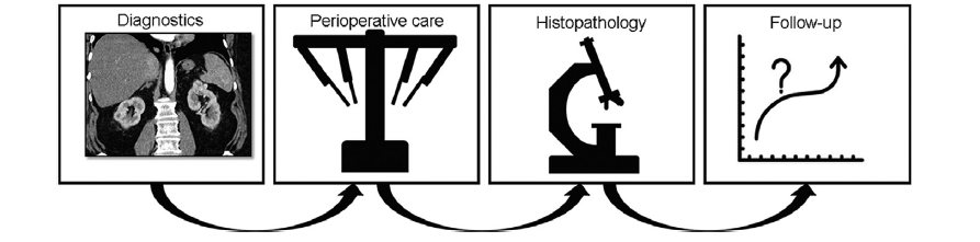
Abstract Artificial intelligence (AI) has made considerable progress within the last decade and is the subject of contemporary literature. This trend is driven by improved computational abilities and increasing amounts of complex data that allow for new approaches in analysis and interpretation. Renal cell carcinoma (RCC) has a rising incidence since most tumors are now detected at an earlier stage due to improved imaging. This creates considerable challenges as approximately 10%-17% of kidney tumors are designated as benign in histopathological evaluation; however, certain co-morbid populations (the obese and elderly) have an increased peri-interventional risk. AI offers an alternative solution by helping to optimize precision and guidance for diagnostic and therapeutic decisions. The narrative review introduced basic principles and provide a comprehensive overview of current AI techniques for RCC. Currently, AI applications can be found in any aspect of RCC management including diagnostics, perioperative care, pathology, and follow-up. Most commonly applied models include neural networks, random forest, support vector machines, and regression. However, for implementation in daily practice, health care providers need to develop a basic understanding and establish interdisciplinary collaborations in order to standardize datasets, define meaningful endpoints, and unify interpretation.
|
|
Received: 30 January 2022
Available online: 20 July 2022
|
|
Corresponding Authors:
Giovanni Cacciamani
E-mail: Giovanni.Cacciamani@med.usc.edu
|
|
|

|
|
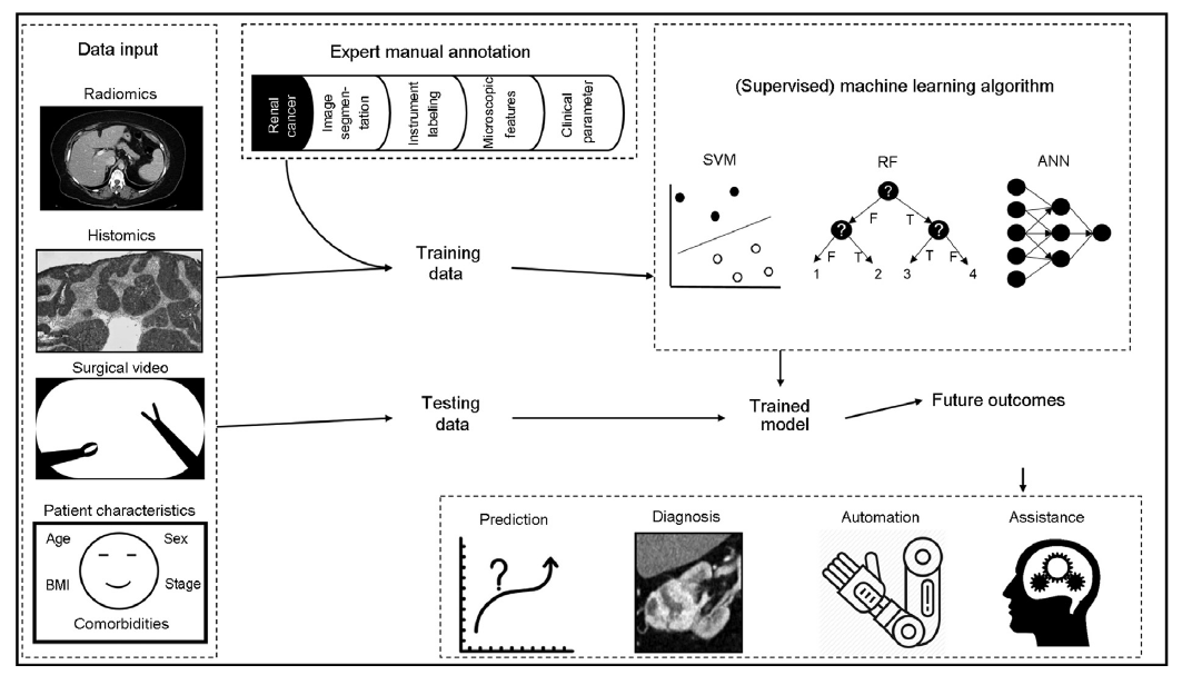
Basic principles of supervised ML models for renal cancer. Available data from different aspects of clinical care can be used as input. Following manual annotation, ML algorithms are trained to create the models. Unused test data are used for validation and to determine the final model which can assist during care of future patients (adopted from Garrow et al. [21]). 1253 mm×714 mm (38×38 DPI). SVM, support vector machine; RF, random forest; ANN, artificial neural networks; ML, machine learning; BMI, body mass index.
|

|
|
Applications of artificial intelligence during the course of treatment. 401 mm×112 mm (38×38 DPI).
|
| [1] |
Ferlay J, Colombet M, Soerjomataram I, Dyba T, Randi G, Bettio M, et al. Cancer incidence and mortality patterns in Europe: estimates for 40 countries and 25 major cancers in 2018. Eur J Cancer 2018; 103:356e87.
doi: S0959-8049(18)30955-9
pmid: 30100160
|
| [2] |
Padala SA, Barsouk A, Thandra KC, Saginala K, Mohammed A, Vakiti A, et al. Epidemiology of renal cell carcinoma. World J Oncol 2020; 11:79e87.
doi: 10.14740/wjon1279
|
| [3] |
Bray F, Ferlay J, Soerjomataram I, Siegel RL, Torre LA, Jemal A. Global cancer statistics 2018: GLOBOCAN estimates of incidence and mortality worldwide for 36 cancers in 185 countries. CA Cancer J Clin 2018; 68:394e424.
doi: 10.3322/caac.21492
|
| [4] |
Ljungberg B, Albiges L, Abu-Ghanem Y, Bensalah K, Dabestani S, Fernandez-Pello S, et al. European Association of Urology guidelines on renal cell carcinoma: the 2019 update. Eur Urol 2019; 75:799e810.
doi: S0302-2838(19)30152-6
pmid: 30803729
|
| [5] |
Moch H, Cubilla AL, Humphrey PA, Reuter VE, Ulbright TM. The 2016 WHO classification of tumours of the urinary system and male genital organsdpart A: renal, penile, and testicular tumours. Eur Urol 2016; 70:93e105.
doi: 10.1016/j.eururo.2016.02.029
|
| [6] |
Han S, Hwang SI, Lee HJ. The classification of renal cancer in 3-phase CT images using a deep learning method. J Digit Imaging 2019; 32:638e43.
doi: 10.1007/s10278-019-00230-2
|
| [7] |
Thorstenson A, Bergman M, Scherman-Plogell AH, Hosseinnia S, Ljungberg B, Adolfsson J, et al. Tumour characteristics and surgical treatment of renal cell carcinoma in Sweden 2005-2010:a population-based study from the national Swedish kidney cancer register. Scand J Urol 2014; 48: 231e8.
doi: 10.3109/21681805.2013.864698
pmid: 24666102
|
| [8] |
Tahbaz R, Schmid M, Merseburger AS. Prevention of kidney cancer incidence and recurrence: lifestyle, medication and nutrition. Curr Opin Urol 2018; 28:62e79.
doi: 10.1097/MOU.0000000000000454
pmid: 29059103
|
| [9] |
Gill IS, Aron M, Gervais DA, Jewett MA. Clinical practice. Small renal mass. N Engl J Med 2010; 362:624e34.
doi: 10.1056/NEJMcp0910041
|
| [10] |
Almassi N, Gill BC, Rini B, Fareed K. Management of the small renal mass. Transl Androl Urol 2017; 6:923e30.
doi: 10.21037/tau.2017.07.11
pmid: 29184793
|
| [11] |
Abou Youssif T, Kassouf W, Steinberg J, Aprikian AG, Laplante MP, Tanguay S. Active surveillance for selected patients with renal masses: updated results with long-term follow-up. Cancer 2007; 110:1010e4.
pmid: 17628489
|
| [12] |
Jewett MA, Mattar K, Basiuk J, Morash CG, Pautler SE, Siemens DR, et al. Active surveillance of small renal masses: progression patterns of early stage kidney cancer. Eur Urol 2011; 60:39e44.
doi: 10.1016/j.eururo.2011.03.030
|
| [13] |
Xu Q, Zhu Q, Liu H, Chang L, Duan S, Dou W, et al. Differentiating benign from malignant renal tumors using T2- and diffusion-weighted images: a comparison of deep learning and radiomics models versus assessment from radiologists. J Magn Reson Imaging 2022; 55:1251e9.
doi: 10.1002/jmri.27900
|
| [14] |
Finelli A, Cheung DC, Al-MatarA, Evans AJ, Morash CG, Pautler SE, et al. Small renal mass surveillance: histology-specific growth rates in a biopsy-characterized cohort. Eur Urol 2020; 78:460e7.
doi: 10.1016/j.eururo.2020.06.053
|
| [15] |
Abel EJ, Culp SH, Matin SF, Tamboli P, Wallace MJ, Jonasch E, et al. Percutaneous biopsy of primary tumor in metastatic renal cell carcinoma to predict high risk pathological features: comparison with nephrectomy assessment. J Urol 2010; 184: 1877e81.
|
| [16] |
Richard PO, Lavallée LT, Pouliot F, Komisarenko M, Martin L, Lattouf JB, et al. Is routine renal tumor biopsy associated with lower rates of benign histology following nephrectomy for small renal masses? J Urol 2018; 200:731e6.
doi: 10.1016/j.juro.2018.04.015
|
| [17] |
Marconi L, Dabestani S, Lam TB, Hofmann F, Stewart F, Norrie J, et al. Systematic review and meta-analysis of diagnostic accuracy of percutaneous renal tumour biopsy. Eur Urol 2016; 69:660e73.
doi: 10.1016/j.eururo.2015.07.072
|
| [18] |
Hameed B, Dhavileswarapu S, Aiswarya V, Raza SZ, Karimi H, Khanuja HS, et al. Artificial intelligence and its impact on urological diseases and management: a comprehensive review of the literature. J Clin Med 2021; 10: 1864e82.
|
| [19] |
Hashimoto DA, Rosman G, Rus D, Meireles OR. Artificial intelligence in surgery: promises and perils. Ann Surg 2018; 268: 70e6.
doi: 10.1097/SLA.0000000000002693
pmid: 29389679
|
| [20] |
Watson DS, Krutzinna J, Bruce IN, Griffiths CE, McInnes IB, Barnes MR, et al. Clinical applications of machine learning algorithms: beyond the black box. BMJ 2019; 364:l886. https://doi.org/10.1136/bmj.l886.
|
| [21] |
Garrow CR, Kowalewski KF, Li L, Wagner M, Schmidt MW, Engelhardt S, et al. Machine learning for surgical phase recognition: a systematic review. Ann Surg 2020; 273:684e93.
doi: 10.1097/SLA.0000000000004425
|
| [22] |
JSC LQA Data annotation for machine learning: A to Z guide. Available from: https://www.lotus-qa.com/data-annotationguide/. [Accessed 30 November 2021].
|
| [23] |
Willemink MJ, Koszek WA, Hardell C, Wu J, Fleischmann D, Harvey H, et al. Preparing medical imaging data for machine learning. Radiology 2020; 295:4e15.
doi: 10.1148/radiol.2020192224
pmid: 32068507
|
| [24] |
Cheng JY, Abel JT, Balis UG, McClintock DS, Pantanowitz L. Challenges in the development, deployment, and regulation of artificial intelligence in anatomic pathology. Am J Pathol 2021; 191:1684e92.
doi: 10.1016/j.ajpath.2020.10.018
|
| [25] |
Mendelson EB. Artificial intelligence in breast imaging: potentials and limitations. AJR Am J Roentgenol 2019; 212:293e9.
|
| [26] |
Xu Y, Goodacre R. On splitting training and validation set: a comparative study of cross-validation, bootstrap and systematic sampling for estimating the generalization performance of supervised learning. J Anal Test 2018; 2:249e62.
doi: 10.1007/s41664-018-0068-2
|
| [27] |
Liaw A, Wiener M. Classification and regression by randomForest. R News 2002; 2:18e22.
|
| [28] |
Formisano E, De Martino F, Valente G. Multivariate analysis of fMRI time series: classification and regression of brain responses using machine learning. Magn Reson Imaging 2008; 26: 921e34.
doi: 10.1016/j.mri.2008.01.052
pmid: 18508219
|
| [29] |
Carmichael I, Marron J. Data science vs. statistics: two cultures? Jpn J Stat Data Sci 2018; 1:117e38.
doi: 10.1007/s42081-018-0009-3
|
| [30] |
Bennett M, Hayes K, Kleczyk EJ, Mehta R. Similarities and differences between machine learning and traditional advanced statistical modeling in healthcare analytics. 2022. arXiv preprint arXiv:2201.02469. https://arxiv.org/abs/2201.02469.
|
| [31] |
Tsili AC, Andriotis E, Gkeli MG, Krokidis M, Stasinopoulou M, Varkarakis IM, et al. The role of imaging in the management of renal masses. Eur J Radiol 2021; 141:109777. https://doi.org/10.1016/j.ejrad.2021.109777.
doi: 10.1016/j.ejrad.2021.109777
|
| [32] |
Kaur R, Juneja M, Mandal AK. An overview of non-invasive imaging modalities for diagnosis of solid and cystic renal lesions. Med Biol Eng Comput 2020; 58:1e24.
doi: 10.1007/s11517-019-02049-z
|
| [33] |
Warshauer DM, McCarthy SM, Street L, Bookbinder MJ, Glickman MG, Richter J, et al. Detection of renal masses: sensitivities and specificities of excretory urography/linear tomography, US, and CT. Radiology 1988; 169:363e5.
|
| [34] |
Hallscheidt PJ, Fink C, Haferkamp A, Bock M, Luburic A, Zuna I, et al. Preoperative staging of renal cell carcinoma with inferior vena cava thrombus using multidetector CT and MRI: prospective study with histopathological correlation. J Comput Assist Tomogr 2005; 29:64e8.
pmid: 15665685
|
| [35] |
Kay FU, Canvasser NE, Xi Y, Pinho DF, Costa DN, Leon AD, et al. Diagnostic performance and interreader agreement of a standardized MR imaging approach in the prediction of small renal mass histology. Radiology 2018; 287:543e53.
doi: 10.1148/radiol.2018171557
|
| [36] |
Cornelis F, Grenier N. Multiparametric magnetic resonance imaging of solid renal tumors: a practical algorithm. Semin Ultrasound CT MR 2017; 38:47e58.
doi: 10.1053/j.sult.2016.08.009
|
| [37] |
Li F, Du L, Xing J, Su Y, Wu Y. Diagnostic efficacy of contrast-enhanced ultrasonography in solid renal parenchymal lesions with maximum diameters of 5 cm. J Ultrasound Med 2008; 27:875e85.
|
| [38] |
Olson MC, Abel EJ, Mankowski Gettle L. Contrast-enhanced ultrasound in renal imaging and intervention. Curr Urol Rep 2019; 20:73. https://doi.org/10.1007/s11934-019-0936-y.
doi: 10.1007/s11934-019-0936-y
|
| [39] |
Tanaka T, Huang Y, Marukawa Y, Tsuboi Y, Masaoka Y, Kojima K, et al. Differentiation of small (_4 cm) renal masses on multiphase contrast-enhanced CT by deep learning. AJR Am J Roentgenol 2020; 214:605e12.
doi: 10.2214/AJR.19.22074
|
| [40] |
Ursprung S, Beer L, Bruining A, Woitek R, Stewart GD, Gallagher FA, et al. Radiomics of computed tomography and magnetic resonance imaging in renal cell carcinomada systematic review and meta-analysis. Eur Radiol 2020; 30: 3558e66.
doi: 10.1007/s00330-020-06666-3
pmid: 32060715
|
| [41] |
Gillies RJ, Kinahan PE, Hricak H. Radiomics: images are more than pictures, they are data. Radiology 2016; 278:563e77.
doi: 10.1148/radiol.2015151169
pmid: 26579733
|
| [42] |
Muhlbauer J, Egen L, Kowalewski KF, Grilli M, Walach MT, Westhoff N, et al. Radiomics in renal cell carcinomada systematic review and meta-analysis. Cancers 2021; 13:1348. https://doi.org/10.3390/cancers13061348.
doi: 10.3390/cancers13061348
|
| [43] |
Ma YC, Xu F, Ma W. Can whole-tumor radiomics-based CT analysis better differentiate fat-poor angiomyolipoma from clear cell renal cell carcinoma: compared with conventional CT analysis? Abdom Radiol (NY) 2020; 45:2500e7.
|
| [44] |
Goh VG, Nathan P, Juttla JK, Vinayan A, Miles KA. Assessment of response to tyrosine kinase inhibitors in metastatic renal cell cancer: CT texture as a predictive biomarker. Radiology 2011; 261:165e71.
doi: 10.1148/radiol.11110264
|
| [45] |
Karimi D, Ruan D. Synergistic combination of learned and hand-crafted features for prostate lesion classification in multiparametric magnetic resonance imaging. In: Descoteaux M, Maier-Hein L, Franz A, Jannin P, Collins D, Duchesne S, editors. Medical image computing and computer assisted interventiondMICCAI 2017, Part III, LNCS 10435. New York:Springer-Verlag; 2017. p.391e8.
|
| [46] |
Cao R, Bajgiran AM, Mirak SA, Shakeri S, Zhong X, Enzmann D, et al. Joint prostate cancer detection and Gleason score prediction in mp-MRI via FocalNet. IEEE Trans Med Imaging 2019; 38:2496e506.
doi: 10.1109/TMI.2019.2901928
|
| [47] |
Chen FG, Hwang D, Cen S, Yap F, Ugwueze C, Varghese B, et al. Voxel-based whole-lesion enhancement parameters: a study of its clinical value in differentiating clear cell renal cell carcinoma from renal oncocytoma. Abdom Radiol (NY) 2017; 42:552e60.
|
| [48] |
Li YH, Xia Y, Long L. Value of radiomics in differential diagnosis of chromophobe renal cell carcinoma and renal oncocytoma. Abdom Radiol (NY) 2020; 45:3193e201.
|
| [49] |
Nassiri N, Maas G, Cacciamani B, Varghese B, Hwang D, Lei X, et al. A radiomic-based machine learning algorithm to reliably differentiate benign renal masses from renal cell carcinoma. Eur Urol Focus 2021; 16:232e7.
|
| [50] |
Qin F, Yuan J. [Research status and trend of artificial intelligence in the diagnosis of urinary diseases]. Sheng Wu Yi Xue Gong Cheng Xue Za Zhi 2020; 37:230e5. [Article in Chinese].
|
| [51] |
Lee M, Wei S, Anaokar J, Uzzo R, Kutikov A. Kidney cancer management 3.0:can artificial intelligence make us better? Curr Opin Urol 2021; 31:409e15.
doi: 10.1097/MOU.0000000000000881
|
| [52] |
Lin F, Cui E, Lei Y, Luo L. CT-based machine learning model to predict the Fuhrman nuclear grade of clear cell renal cell carcinoma. Abdom Radiol (NY) 2019; 44:2528e34.
|
| [53] |
Nazari M, Shiri I, Zaidi H. Radiomics-based machine learning model to predict risk of death within 5-years in clear cell renal cell carcinoma patients. Comput Biol Med 2021; 129:104135. https://doi.org/10.1016/j.compbiomed.2020.104135.
doi: 10.1016/j.compbiomed.2020.104135
|
| [54] |
Kocak B, Kaya OK, Erdim C, Kus EA, Kilickesmez O. Artificial intelligence in renal mass characterization: a systematic review of methodologic items related to modeling, performance evaluation, clinical utility, and transparency. AJR Am J Roentgenol 2020; 215:1113e22.
doi: 10.2214/AJR.20.22847
|
| [55] |
Doyle PW, Kavoussi NL. Machine learning applications to enhance patient specific care for urologic surgery. World J Urol 2021; 40:679e86.
doi: 10.1007/s00345-021-03738-x
|
| [56] |
Kowalewski KF, Garrow CR, Schmidt MW, Benner L, MÜller-Stich BP, Nickel F. Sensor-based machine learning for workflow detection and as key to detect expert level in laparoscopic suturing and knot-tying. Surg Endosc 2019; 33:3732e40.
doi: 10.1007/s00464-019-06667-4
|
| [57] |
Kowalewski KF, Hendrie JD, Schmidt MW, Garrow CR, Bruckner T, Proctor T, et al. Development and validation of a sensor- and expert model-based training system for laparoscopic surgery: the iSurgeon. Surg Endosc 2017; 31: 2155e65.
|
| [58] |
Nakawala H, Bianchi R, Pescatori LE, De Cobelli O, Ferrigno G, De Momi E. “Deep-Onto” network for surgical workflow and context recognition. Int J Comput Assist Radiol Surg 2019; 14: 685e96.
doi: 10.1007/s11548-018-1882-8
|
| [59] |
Zhao B, Waterman RS, Urman RD, Gabriel RA. A machine learning approach to predicting case duration for robot-assisted surgery. J Med Syst 2019; 43:32. https://doi.org/10.1007/s10916-018-1151-y.
doi: 10.1007/s10916-018-1151-y
|
| [60] |
Bhandari M, Nallabasannagari AR, Reddiboina M, Porter JR, Jeong W, Mottrie A, et al. Predicting intra-operative and postoperative consequential events using machine-learning techniques in patients undergoing robot-assisted partial nephrectomy: a Vattikuti Collective Quality Initiative database study. BJU Int 2020; 126:350e8.
|
| [61] |
Ross T, Zimmerer D, Vemuri A, Isensee F, Wiesenfarth M, Bodenstedt S, et al. Exploiting the potential of unlabeled endoscopic video data with self-supervised learning. Int J Comput Assist Radiol Surg 2018; 13:925e33.
doi: 10.1007/s11548-018-1772-0
|
| [62] |
Hung AJ, Chen J, Che Z, Nilanon T, Jarc A, Titus M, et al. Utilizing machine learning and automated performance metrics to evaluate robot-assisted radical prostatectomy performance and predict outcomes. J Endourol 2018; 32:438e44.
doi: 10.1089/end.2018.0035
|
| [63] |
Hung AJ, Chen J, Gill IS. Automated performance metrics and machine learning algorithms to measure surgeon performance and anticipate clinical outcomes in robotic surgery. JAMA Surg 2018; 153:770e1.
doi: 10.1001/jamasurg.2018.1512
|
| [64] |
Ghodoussipour S, Reddy SS, Ma R, Huang D, Nguyen J, Hung AJ. An objective assessment of performance during robotic partial nephrectomy: validation and correlation of automated performance metrics with intraoperative outcomes. J Urol 2021; 205:1294e302.
doi: 10.1097/JU.0000000000001557
pmid: 33356480
|
| [65] |
Amparore D, Pecoraro A, Checcucci E, Piramide F, Verri P, De Cillis S, et al. Three-dimensional virtual models’ assistance during minimally invasive partial nephrectomy minimizes the impairment of kidney function. Eur Urol Oncol 2022; 5:104e8.
|
| [66] |
Schiavina R, Bianchi L, Chessa F, Barbaresi U, Cercenelli L, Lodi S, et al. Augmented reality to guide selective clamping and tumor dissection during robot-assisted partial nephrectomy: a preliminary experience. Clin Genitourin Cancer 2021; 19:e149e55. https://doi.org/10.1016/j.clgc.2020.09.005.
doi: 10.1016/j.clgc.2020.09.005
|
| [67] |
Porpiglia F, Checcucci E, Amparore D, Piramide F, Volpi G, Granato S, et al. Three-dimensional augmented reality robot-assisted partial nephrectomy in case of complex tumours (PADUA_10):a new intraoperative tool overcoming the ultrasound guidance. Eur Urol 2020; 78:229e38.
doi: 10.1016/j.eururo.2019.11.024
|
| [68] |
Nosrati MS, Amir-Khalili A, Peyrat JM, Abinahed J, Al-Alao O, Al-Ansari A, et al. Endoscopic scene labelling and augmentation using intraoperative pulsatile motion and colour appearance cues with preoperative anatomical priors. Int J Comput Assist Radiol Surg 2016; 11:1409e18.
doi: 10.1007/s11548-015-1331-x
pmid: 26872810
|
| [69] |
Amir-Khalili A, Hamarneh G, Peyrat JM, Abinahed J, Al-Alao O, Al-Ansari A, et al. Automatic segmentation of occluded vasculature via pulsatile motion analysis in endoscopic robot-assisted partial nephrectomy video. Med Image Anal 2015; 25:103e10.
doi: 10.1016/j.media.2015.04.010
pmid: 25977157
|
| [70] |
Haifler M, Pence I, Sun Y, Kutikov A, Uzzo RG, Mahadevan A, et al. Discrimination of malignant and normal kidney tissue with short wave infrared dispersive Raman spectroscopy. J Biophotonics 2018; 11:e201700188. https://doi.org/10.1002/jbio.201700188.
|
| [71] |
Nagpal K, Foote D, Liu Y, Chen PC, Wulczyn E, Tan F, et al. Development and validation of a deep learning algorithm for improving Gleason scoring of prostate cancer. NPJ Digit Med 2019; 2:48. https://doi.org/10.1038/s41746-019-0112-2.
doi: 10.1038/s41746-019-0112-2
pmid: 31304394
|
| [72] |
Wessels F, Schmitt M, Krieghoff-Henning E, Jutzi T, Worst TS, Waldbillig F, et al. Deep learning approach to predict lymph node metastasis directly from primary tumour histology in prostate cancer. BJU Int 2021; 128:352e60.
doi: 10.1111/bju.15386
|
| [73] |
Holdbrook DA, Singh M, Choudhury Y, Kalaw EM, Koh V, Tan H, et al. Automated renal cancer grading using nuclear pleomorphic patterns. JCO Clin Cancer Inform 2018; 2:1e12.
doi: 10.1200/CCI.17.00100
pmid: 30652593
|
| [74] |
Tabibu S, Vinod P, Jawahar C. Pan-renal cell carcinoma classification and survival prediction from histopathology images using deep learning. Sci Rep 2019; 9:1e9.
doi: 10.1038/s41598-018-37186-2
|
| [75] |
Tian K, Rubadue CA, Lin DI, Veta M, Pyle ME, Irshad H, et al. Automated clear cell renal carcinoma grade classification with prognostic significance. PLoS One 2019; 14:e0222641. https://doi.org/10.1371/journal.pone.0222641.
|
| [76] |
Fenstermaker M, Tomlins SA, Singh K, Wiens J, Morgan TM. Development and validation of a deep-learning model to assist with renal cell carcinoma histopathologic interpretation. Urology 2020; 144:152e7.
doi: S0090-4295(20)30856-6
pmid: 32711010
|
| [77] |
Yeh FC, Parwani AV, Pantanowitz L, Ho C. Automated grading of renal cell carcinoma using whole slide imaging. J Pathol Inform 2014; 5:23. https://doi.org/10.4103/2153-3539.137726.
doi: 10.4103/2153-3539.137726
|
| [78] |
Khoshdeli M, Borowsky A, Parvin B. Deep learning models differentiate tumor grades from H&E stained histology sections. Annu Int Conf IEEE Eng Med Biol Soc 2018; 2018:620e3.
|
| [79] |
He Z, Liu H, Moch H, Simon HU. Machine learning with autophagy-related proteins for discriminating renal cell carcinoma subtypes. Sci Rep 2020; 10:720. https://doi.org/10.1038/s41598-020-57670-y.
doi: 10.1038/s41598-020-57670-y
|
| [80] |
Singh NP, Bapi RS, Vinod PK. Machine learning models to predict the progression from early to late stages of papillary renal cell carcinoma. Comput Biol Med 2018; 100:92e9.
|
| [81] |
Singh NP, Vinod P. Integrative analysis of DNA methylation and gene expression in papillary renal cell carcinoma. Mol Genet Genomics 2020; 295:807e24.
|
| [82] |
Brennan K, Metzner TJ, Kao CS, Massie CE, Stewart GD, Haile RW, et al. Development of a DNA methylationdbased diagnostic signature to distinguish benign oncocytoma from renal cell carcinoma. JCO Precis Oncol 2020; 4:1141e51.
|
| [83] |
Hung AJ, Chen J, Ghodoussipour S, Oh PJ, Liu Z, Nguyen J, et al. Deep learning on automated performance metrics and clinical features to predict urinary continence recovery after robot-assisted radical prostatectomy. BJU Int 2019;124.
|
| [84] |
Hung AJ, Chen J, Jarc A, Hatcher D, Djaladat H, Gill IS. Development and validation of objective performance metrics for robot-assisted radical prostatectomy: a pilot study. J Urol 2018; 199:296e304.
doi: 10.1016/j.juro.2017.07.081
|
| [85] |
Katzman JL, Shaham U, Cloninger A, Bates J, Jiang T, Kluger Y. DeepSurv: personalized treatment recommender system using a Cox proportional hazards deep neural network. BMC Med Res Methodol 2018; 18:24. https://doi.org/10.1186/s12874-018-0482-1.
doi: 10.1186/s12874-018-0482-1
pmid: 29482517
|
| [86] |
Wong NC, Lam C, Patterson L, Shayegan B. Use of machine learning to predict early biochemical recurrence after robot-assisted prostatectomy. BJU Int 2019; 123:51e7.
doi: 10.1111/bju.14477
|
| [87] |
Kattan MW. Comparison of Cox regression with other methods for determining prediction models and nomograms. J Urol 2003; 170:S6e10.
|
| [88] |
kattan MW, Reuter V, Motzer RJ, Katz J, Russo P. A postoperative prognostic nomogram for renal cell carcinoma. J Urol 2001; 166:63e7.
doi: 10.1016/S0022-5347(05)66077-6
|
| [89] |
Kim H, Lee SJ, Park SJ, Choi IY, Hong SH. Machine learning approach to predict the probability of recurrence of renal cell carcinoma after surgery: prediction model development study. JMIR Med Informatics 2021; 9:e25635. https://doi.org/10.2196/25635.
doi: 10.2196/25635
|
| [90] |
Guo Y, Braga L, Kapoor A. PD07-08machine learning to predict recurrence of localized renal cell carcinoma. J Urol 2019; 201: e145. https://doi.org/10.1097/01.JU.0000555241.27498.f6.
|
| [91] |
Brodie A, Dai N, Teoh YCJ, Decaestecker K, Dasgupta P, Vasdev N. Artificial intelligence in urological oncology: an update and future applications. Urol Oncol 2021; 39: 379e99.
doi: 10.1016/j.urolonc.2021.03.012
|
| [92] |
Buchner A, Kendlbacher M, Nuhn P, TÜllmann C, Haseke N, Stief CG, et al. Outcome assessment of patients with metastatic renal cell carcinoma under systemic therapy using artificial neural networks. Clin Genitourin Cancer 2012; 10:37e42.
doi: 10.1016/j.clgc.2011.10.001
|
| [1] |
Stefano Puliatti,Ahmed Eissa,Enrico Checcucci,Pietro Piazza,Marco Amato,Stefania Ferretti,Simone Scarcella,Juan Gomez Rivas,Mark Taratkin,Josè Marenco,Ines Belenchon Rivero,Karl-Friedrich Kowalewski,Giovanni Cacciamani,Ahmed El-Sherbiny,Ahmed Zoeir,Abdelhamid M. El-Bahnasy,Ruben De Groote,Alexandre Mottrie,Salvatore Micali. New imaging technologies for robotic kidney cancer surgery[J]. Asian Journal of Urology, 2022, 9(3): 253-262. |
| [2] |
Daniele Amparore,Angela Pecoraro,Federico Piramide,Paolo Verri,Enrico Checcucci,Sabrina De Cillis,Alberto Piana,Mariano Burgio,Michele Di Dio,Matteo Manfredi,Cristian Fiori,Francesco Porpiglia. Three-dimensional imaging reconstruction of the kidney's anatomy for a tailored minimally invasive partial nephrectomy: A pilot study[J]. Asian Journal of Urology, 2022, 9(3): 263-271. |
| [3] |
Luke P. O’Connor,Shayann Ramedani,Michael Daneshvar,Arvin K. George,Andre Luis Abreu,Giovanni E. Cacciamani,Amir H. Lebastchi. Future perspective of focal therapy for localized prostate cancer[J]. Asian Journal of Urology, 2021, 8(4): 354-361. |
| [4] |
Shuchi Gulati,Nicholas J. Vogelzang. Biomarkers in renal cell carcinoma: Are we there yet?[J]. Asian Journal of Urology, 2021, 8(4): 362-375. |
| [5] |
Kulthe Ramesh Seetharam Bhat,Srinivas Samavedi,Marcio Covas Moschovas,Fikret Fatih Onol,Shannon Roof,Travis Rogers,Vipul R. Patel,Ananthakrishnan Sivaraman. Magnetic resonance imaging-guided prostate biopsy—A review of literature[J]. Asian Journal of Urology, 2021, 8(1): 105-116. |
| [6] |
Yucong Zhang,Gongwei Long,Haojie Shang,Beichen Ding,Guoliang Sun,Wei Ouyang,Man Liu,Yuan Chen,Heng Li,Hua Xu,Zhangqun Ye. Comparison of the oncological, perioperative and functional outcomes of partial nephrectomy versus radical nephrectomy for clinical T1b renal cell carcinoma: A systematic review and meta-analysis of retrospective studies[J]. Asian Journal of Urology, 2021, 8(1): 117-125. |
| [7] |
lIlan Klein,Jorge Gutiérrez-Aceves. Preoperative imaging in staghorn calculi, planning and decision making in management of staghorn calculi[J]. Asian Journal of Urology, 2020, 7(2): 87-93. |
| [8] |
Bohdan Baralo,Patrick Samson,David Hoenig,Arthur Smith. Percutaneous kidney stone surgery and radiation exposure: A review[J]. Asian Journal of Urology, 2020, 7(1): 10-17. |
| [9] |
Brian T. Hanyok,Mary M. Everist,Lauren E. Howard,Amanda M. De Hoedt,William J. Aronson,Matthew R. Cooperberg,Christopher J. Kane,Christopher L. Amling,Martha K. Terris,Stephen J. Freedland. Practice patterns and outcomes of equivocal bone scans for patients with castration-resistant prostate cancer: Results from SEARCH[J]. Asian Journal of Urology, 2019, 6(3): 242-248. |
| [10] |
Edwin Jonathan Aslim,Yan Mee Law,Puay Hoon Tan,John Carson Allen Jr,Lionel Tim-Ee Cheng,Viswanath Anand Chidambaram,Li Yan Khor,Benjamin Yongcheng Tan,Ernest Wencong Eu,Christopher Wai Sam Cheng,John Shyi Peng Yuen,Henry Sun Sien Ho,Lui Shiong Lee. Multiparametric MRI reporting using Prostate Imaging Reporting and Data System version 2.0 (PI-RADSv2) retains clinical efficacy in a predominantly post-biopsy patient population[J]. Asian Journal of Urology, 2019, 6(3): 256-263. |
| [11] |
Jean-Luc Descotes. Diagnosis of prostate cancer[J]. Asian Journal of Urology, 2019, 6(2): 129-136. |
| [12] |
Olivier Rouviere,Paul Cezar Moldovan. The current role of prostate multiparametric magnetic resonance imaging[J]. Asian Journal of Urology, 2019, 6(2): 137-145. |
| [13] |
Kenneth Chen,Kae Jack Tay,Yan Mee Law,Hakan Aydin,Henry Ho,Christopher Cheng,John Shyi Peng Yuen. Outcomes of combination MRI-targeted and transperineal template biopsy in restaging low-risk prostate cancer for active surveillance[J]. Asian Journal of Urology, 2018, 5(3): 184-193. |
| [14] |
Kai Zhang, Chris H. Bangma, Monique J. Roobol. Prostate cancer screening in Europe and Asia[J]. Asian Journal of Urology, 2017, 4(2): 86-95. |
| [15] |
Geoffrey S. Gaunay, Vinay Patel, Paras Shah, Daniel Moreira, Ardeshir R. Rastinehad, Eran Ben-Levi, Robert Villani, Manish A. Vira. Multi-parametric MRI of the prostate: Factors predicting extracapsular extension at the time of radical prostatectomy[J]. Asian Journal of Urology, 2017, 4(1): 31-36. |
|
|
|
|