Color Doppler ultrasound imaging in varicoceles: Is the difference in venous diameter encountered during Valsalva predictive of palpable varicocele grade?
Kelly Lehner*(),Catherine Ingram,Utsav Bansal,Colleen Baca,Adithya Balasubramanian,Nannan Thirumavalavan,Jason M. Scovell,Saneal Rajanahally,Matthew Pollard,Larry I. Lipshultz
Baylor College of Medicine, Scott Department of Urology, Houston, TX, USA
Objective: The clinical grading system for varicoceles is subjective and dependent on clinician experience. Color Doppler ultrasound (US) has not been standardized in the diagnosis of varicoceles. We aimed to determine if US measurement of varicocele could be predictive of World Health Organization (WHO) varicocele grade.
Methods: Men who presented for either scrotal pain or infertility to a tertiary men's health clinic underwent physical examination, and varicoceles were graded following WHO criteria (0=subclinical, 1, 2, 3). US was used to measure largest venous diameter in the pampiniform plexus bilaterally at rest and during Valsalva maneuver. Receiver operator characteristic curve analysis was used to determine if resting diameter, diameter during Valsalva, or change in diameter between at rest and during Valsalva provided the highest sensitivity and specificity for determining clinical grade. Threshold values for diameter were determined from these receiver operator characteristic curves.
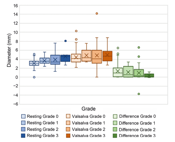
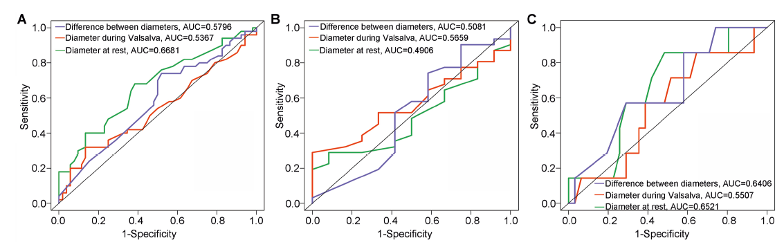
Results: A total of 102 men (50 with clinical varicocele and 52 with subclinical varicocele) were included. Diameter at rest was the best ultrasonographic discriminator between subclinical and clinical varicoceles (area under the curve [AUC]=0.67) with a diameter threshold of 3.0 mm (sensitivity 79%, specificity 42%). Diameter during Valsalva had the greatest AUC for determining clinical Grades 1 versus 2 (AUC=0.57) with diameter threshold of 5.7 mm (sensitivity 71%, specificity 33%). For differentiating between Grades 2 and 3, diameter at rest had the greatest AUC of 0.65 with a threshold of 3.6 mm (sensitivity 71%, specificity 58%).
Conclusion: Our results corroborate other studies that have shown a weak correlation between US and clinical grading. The use of diameter during Valsalva was less predictive than diameter at rest and was only clinically significant in differentiating between Grade 1 and 2 varicocele. A standardized method for determining clinically relevant varicoceles on US would allow for improved patient counseling and clinical decision-making.
. [J]. Asian Journal of Urology, 2023, 10(1): 27-32.
Kelly Lehner,Catherine Ingram,Utsav Bansal,Colleen Baca,Adithya Balasubramanian,Nannan Thirumavalavan,Jason M. Scovell,Saneal Rajanahally,Matthew Pollard,Larry I. Lipshultz. Color Doppler ultrasound imaging in varicoceles: Is the difference in venous diameter encountered during Valsalva predictive of palpable varicocele grade?. Asian Journal of Urology, 2023, 10(1): 27-32.
Paick S, Choi WS. Varicocele and testicular pain: a review. World J Mens Health 2019; 37:4e11.
doi: 10.5534/wjmh.170010
pmid: 29774668
[2]
American Urological Association Education and Research, Inc. Report on varicocele and infertility: an AUA best practice policy and ASRM practice committee report https://www.auanet.org/documents/education/clinical-guidance/Varicocele-Archive.pdf. [Accessed 1 July 2021].
[3]
Gat Y, Bachar GN, Zukerman Z, Belenky A, Gorenish M. Phys-ical examination may miss the diagnosis of bilateral varico-cele: a comparative study of 4 diagnostic modalities. J Urol 2004; 172:1414e7.
doi: 10.1097/01.ju.0000138540.57137.5f
[4]
Jungwirth A, Giwercman A, Tournaye H, Diemer T, Kopa Z, Dohle G, et al. European association of Urology guidelines on male infertility: the 2012 update. Eur Urol 2012; 62:324e32.
doi: 10.1016/j.eururo.2012.04.048
pmid: 22591628
[5]
Dubin L, Amelar RD. Varicocele size and results of varicoce-lectomy in selected subfertile men with varicocele. Fertil Steril 1970; 21:606e9.
doi: 10.1016/s0015-0282(16)37684-1
pmid: 5433164
[6]
Rowe PJ, Comhaire FH, Mahmoud AM. WHO manual for the standardized investigation, diagnosis, and management of the infertile male. https://apps.who.int/iris/handle/10665/42437. [Accessed 27 November 2021].
[7]
Freeman S, Bertolotto M, Richenberg J, Bel?eld J, Dogra V, Huang DY, et al. Ultrasound evaluation of varicoceles: guidelines and recommendations of the European Society of Urogenital Radiology Scrotal and Penile Imaging Working Group (ESUR-SPIWG) for detection, classi?cation, and grading. Eur Radiol 2020; 30:11e25.
doi: 10.1007/s00330-019-06280-y
pmid: 31332561
[8]
Cocuzza MS, Tiseo BC, Srougi V, Wood GJA, Cardoso JPGF, Esteves SC, et al. Diagnostic accuracy of physical examination compared with color Doppler ultrasound in the determination of varicocele diagnosis and grading: impact of urologists’ experience. Andrology 2020; 8:1160e6.
doi: 10.1111/andr.12797
[9]
World Health Organization. The influence of varicocele on parameters of fertility in a large group of men presenting to infertility clinics. World health organization. Fertil Steril 1992; 57:1289e93.
pmid: 1601152
[10]
Hadziselimovic F, Herzog B, Jenny P. The chance for fertility in adolescent boys after corrective surgery for varicocele. J Urol 1995; 154:731e3.
doi: 10.1097/00005392-199508000-00106
pmid: 7609165
[11]
Thirumavalavan N, Scovell JM, Balasubramanian A, Kohn TP, Ji B, Hasan A, et al. The impact of microsurgical repair of subclinical and clinical varicoceles on total motile sperm count: is there a difference? Urology 2018; 120:109e13.
doi: S0090-4295(18)30627-7
pmid: 29981299
Al-Ali BM, Marszalek M, Shamloul R, Pummer K, Trummer H. Clinical parameters and semen analysis in 716 Austrian pa-tients with varicocele. Urology 2010; 75:1069e73.
doi: 10.1016/j.urology.2009.11.042
[14]
Grasso M, Lania C, Castelli M, Galli L, Franzoso F, Rigatti P. Low-grade left varicocele in patients over 30 years old: the effect of spermatic vein ligation on fertility. BJU Int 2000; 85:305e7.
pmid: 10671887
[15]
Samplaski MK, Jarvi KA. Prognostic factors for a favorable outcome after varicocele repair in adolescents and adults. Asian J Androl 2016; 18:217e21.
doi: 10.4103/1008-682X.169558
pmid: 26732108
[16]
Ishikawa T, Fujisawa M. Effect of age and grade on surgery for patients with varicocele. Urology 2005; 65:768e72.
pmid: 15833525
[17]
Takahara M, Ichikawa T, Shiseki Y, Nakamura T, Shimazaki J. Relationship between grade of varicocele and the response to varicocelectomy. Int J Urol 1996; 3:282e5.
pmid: 8844284
[18]
Steckel J, Dicker AP, Goldstein M. Relationship between varicocele size and response to varicocelectomy. J Urol 1993; 149:769e71.
doi: 10.1016/s0022-5347(17)36203-1
pmid: 8455240
[19]
Orda R, Sayfan J, Manor H, Witz E, Sofer Y. Diagnosis of varicocele and postoperative evaluation using inguinal ultra-sonography. Ann Surg 1987; 206:99e101.
pmid: 3300579
[20]
Leslie SW, Sajjad H, Siref LE. Varicocele. In: StatPearls [Internet]. Treasure Island (FL): StatPearls Publishing; 2020. https://www.ncbi.nlm.nih.gov/books/NBK448113/.[Accessed 1 July 2021].
[21]
Caskurlu T, Tasci AI, Resim S, Sahinkanat T, Ekerbicer H. Reliability of venous diameter in the diagnosis of subclinical varicocele. Urol Int 2003; 71:83e6.
pmid: 12845267
[22]
Cina A, Minnetti M, Pirronti T, Spampinato MV, Canadè A, Oliva G, et al. Sonographic quantitative evaluation of scrotal veins in healthy subjects: normative values and implications for the diagnosis of varicocele. Eur Urol 2006; 50:345e50.
pmid: 16542771
[23]
Hoekstra T, Witt MA. The correlation of internal spermatic vein palpability with ultrasonographic diameter and reversal of venous flow. J Urol 1995; 153:82e4.
doi: 10.1097/00005392-199501000-00029
pmid: 7966798
[24]
Metin A, Bulut O, Temizkan M. Relationship between the left spermatic vein diameter measured by ultrasound and palpated varicocele and Doppler ultrasound ?ndings. Int Urol Nephrol 1991; 23:65e8.
pmid: 1938219
[25]
Pilatz A, Altinkilic B, Kohler E, Marconi M, Weidner W. Color Doppler ultrasound imaging in varicoceles: is the venous diameter suf?cient for predicting clinical and subclinical varicocele? World J Urol 2011; 29:645e50.
doi: 10.1007/s00345-011-0701-4
pmid: 21607575
[26]
Karami M, Mazdak H, Khanbabapour S, Adibi A, Nasr N. Determination of the best position and site for color Doppler ultrasonographic evaluation of the testicular vein to de?ne the clinical grades of varicocele ultrasonographically. Adv Biomed Res 2014; 3:17. https://doi.org/10.4103/2277-9175.124647.
doi: 10.4103/2277-9175.124647
pmid: 24592367