aDepartment of Urology, the First Affiliated Hospital of Nanjing Medical University, Nanjing, China bNanjing Hospital of Traditional Chinese Medicine, Nanjing Hospital of Chinese Medicine, Nanjing, China cDepartment of Urology, Wuhu Hospital Affiliated to East China Normal University, Wuhu, China dDepartment of Urology, Suzhou Ninth People's Hospital, Soochow University, Suzhou, China eDepartment of Laboratory, the First Affiliated Hospital of Nanjing Medical University, Nanjing, China
Objective: This study aimed to analyze the correlation between urinary crystals and urinary calculi.
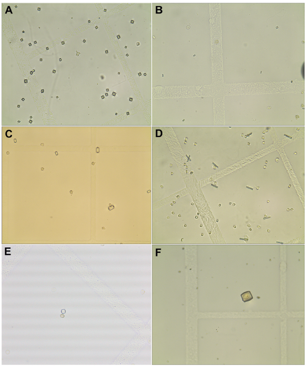
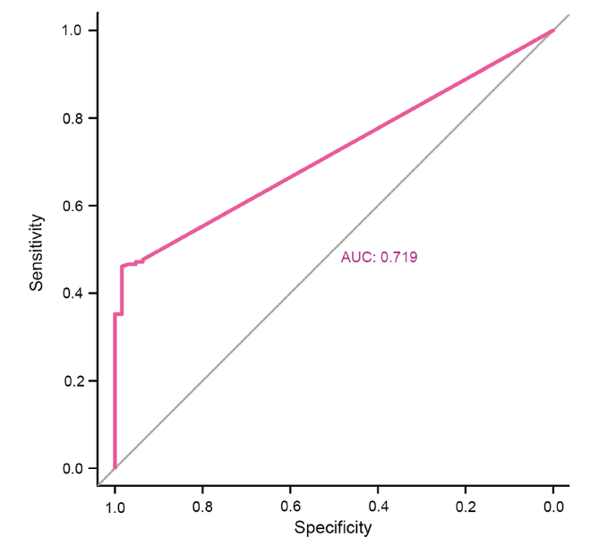
Methods: Clinical data, including urinary crystal types, were collected from 237 patients with urinary calculi. The detection rate of urine crystals and their correlation with stone composition were analyzed. The receiver operating characteristic curve analysis was used to determine the best cut-off value for predicting stone formation risk based on calcium oxalate crystals in urine.
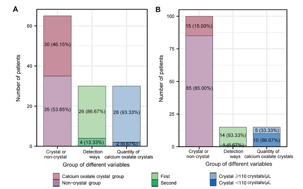
Results: Calcium oxalate was the most common component in 237 patients. Among them, 201 (84.81%) patients had stones containing calcium oxalate. In these patients, calcium oxalate crystals were detected in 45.77% (92/201) of cases. In different groups of calcium oxalate stones, calcium oxalate crystals accounted for more than 90% of the total number of crystals detected in each group. The detection rate of calcium oxalate crystals was higher in first-time stone formers than in recurrent patients. The receiver operating characteristic curve analysis suggested a cut-off value of 110 crystals/μL for predicting stone formation, validated with 65 patients and 100 normal people.
Conclusion: Calcium oxalate crystals in urine can predict the composition of calcium oxalate stones and indicate a higher risk of stone formation when the number exceeds 110 crystals/μL. This non-invasive method may guide clinical treatment and prevention strategies.
Scales CD, Smith AC, Hanley JM, Saigal CS. Prevalence of kidney stones in the United States. Eur Urol 2012; 62:160-5.
doi: 10.1016/j.eururo.2012.03.052
pmid: 22498635
[2]
Wang W, Fan J, Huang G, Li J, Zhu X, Tian Y, et al. Prevalence of kidney stones in mainland China: a systematic review. Sci Rep 2017; 7:41630. https://doi.org/10.1038/srep41630.
doi: 10.1038/srep41630
pmid: 28139722
[3]
Zeng G, Mai Z, Xia S, Wang Z, Zhang K, Wang L, et al. Prevalence of kidney stones in China: an ultrasonography based cross-sectional study. BJU Int 2017; 120:109-16.
doi: 10.1111/bju.13828
pmid: 28236332
[4]
Alexander RT, Hemmelgarn BR, Wiebe N, Bello A, Morgan C, Samuel S, et al. Kidney stones and kidney function loss: a cohort study. BMJ 2012; 345:e5287. https://doi.org/10.1136/bmj.e5287.
Singh P, Enders FT, Vaughan LE, Bergstralh EJ, Knoedler JJ, Krambeck AE, et al. Stone composition among first-time symptomatic kidney stone formers in the community. Mayo Clin Proc 2015; 90:1356-65.
doi: 10.1016/j.mayocp.2015.07.016
pmid: 26349951
[7]
Worcester EM, Coe FL. Clinical practice. Calcium kidney stones. N Engl J Med 2010; 363:954-63.
[8]
Kok DJ, Boellaard W, Ridwan Y, Levchenko VA. Timelines of the “free-particle” and “fixed-particle” models of stoneformation: theoretical and experimental investigations. Urolithiasis 2017; 45:33-41.
doi: 10.1007/s00240-016-0946-x
pmid: 27915394
[9]
Finlayson B. Physicochemical aspects of urolithiasis. Kidney Int 1978; 13:344-60.
pmid: 351263
[10]
Frochot V, Daudon M. Clinical value of crystalluria and quantitative morphoconstitutional analysis of urinary calculi. Int J Surg 2016; 36:624-32.
doi: S1743-9191(16)31032-9
pmid: 27847293
[11]
Robert M, Boularan AM, Delbos O, Guiter J, Descomps B. Study of calcium oxalate crystalluria on renal and vesical urines in stone formers and normal subjects. Urol Int 1998; 60:41-6.
pmid: 9519420
[12]
Fink HA, Wilt TJ, Eidman KE, Garimella PS, MacDonald R, Rutks IR, et al. Medical management to prevent recurrent nephrolithiasis in adults: a systematic review for an American College of Physicians clinical guideline. Ann Intern Med 2013; 158:535-43.
doi: 10.7326/0003-4819-158-7-201304020-00005
pmid: 23546565
[13]
Brikowski TH, Lotan Y, Pearle MS. Climate-related increase in the prevalence of urolithiasis in the United States. Proc Natl Acad Sci U S A 2008; 105:9841-6.
[14]
Ye Z, Zeng G, Yang H, Li J, Tang K, Wang G, et al. The status and characteristics of urinary stone composition in China. BJU Int 2020; 125:801-9.
doi: 10.1111/bju.14765
pmid: 30958622
[15]
Burns JR, Finlayson B, Gauthier J. Calcium oxalate retention in subjects with crystalluria. Urol Int 1984; 39:36-9.
pmid: 6730116
[16]
Robertson WG. Potential role of fluctuations in the composition of renal tubular fluid through the nephron in the initiation of Randall’s plugs and calcium oxalate crystalluria in a computer model of renal function. Urolithiasis 2015; 43(Suppl 1): 93-107. https://doi.org/10.1007/s00240-014-0737-1.
Lieske JC, Leonard R, Toback FG. Adhesion of calcium oxalate monohydrate crystals to renal epithelial cells is inhibited by specific anions. Am J Physiol 1995; 268:F604-12. https://doi.org/10.1152/ajprenal.1995.268.4.F604.
[20]
Gill WB, Jones KW, Ruggiero KJ. Protective effects of heparin and other sulfated glycosaminoglycans on crystal adhesion to injured urothelium. J Urol 1982; 127:152-4.
doi: 10.1016/s0022-5347(17)53650-2
pmid: 6173491
[21]
Cavanaugh C, Perazella MA. Urine sediment examination in the diagnosis and management of kidney disease: core curriculum 2019. Am J Kidney Dis 2019; 73:258-72.
doi: S0272-6386(18)30873-4
pmid: 30249419
[22]
Berg W, Schnapp JD, Schneider HJ, Hesse A, Hienzsch E. Crystaloptical and spectroscopical findings with calcium oxalate crystals in the urine sediment: a contribution to the genesis of oxalate stones. Eur Urol 1976; 2:92-7.
pmid: 971679
[23]
Azoury R, Garside J, Robertson WG. Calcium oxalate precipitation in a flow system: an attempt to simulate the early stages of stone formation in the renal tubules. J Urol 1986; 136:150-3.
doi: 10.1016/s0022-5347(17)44761-6
pmid: 3712603
[24]
Robertson WG, Peacock M, Nordin BE. Calcium crystalluria in recurrent renal-stone formers. Lancet 1969; 2:21-4.
doi: 10.1016/s0140-6736(69)92598-7
pmid: 4182793
[25]
Daudon M, Hennequin C, Boujelben G, Lacour B, Jungers P. Serial crystalluria determination and the risk of recurrence in calcium stone formers. Kidney Int 2005; 67:1934-43.
pmid: 15840041
Luis G. Medina, Randall A. Lee, Valeria Celis, Veronica Rodriguez, Jaime Poncel, Aref S. Sayegh, Rene Sotelo. Robotic management of urinary fistula[J]. Asian Journal of Urology, 2024, 11(3): 357
-365
.