|
|
|
| The dual pathogenesis of penile neoplasia: The heterogeneous morphology of human papillomavirus-related tumors |
Alcides Chauxa,Diego F. Sanchezb,María José Fernández-Nestosac,Sofía Cañete-Portillod,Ingrid M. Rodríguezb,e,Giovanna A. Giannicof,Antonio L. Cubillab,e,*( ) )
|
aDepartment of Scientific Research, School of Postgraduate Studies Norte University, Asunción, Paraguay
bInstituto de Patología e Investigación, Asunción, Paraguay
cPolytechnic School, National University of Asunción, San Lorenzo, Paraguay
dDepartment of Pathology, University of Alabama at Birmingham, Birmingham, AL, USA
eSchool of Medicine, National University of Asunción, San Lorenzo, Paraguay
fDepartment of Pathology, Microbiology, and Immunology, Vanderbilt University Medical Center, Nashville, TN, USA |
|
|
|
|
Abstract Objective: Penile neoplasia, usually of squamous histogenesis, is currently classified into human papillomavirus (HPV)-related or -dependent and non-HPV-related or -independent. There are distinct morphological differences among the two groups. New research studies on penile cancer from Northern countries showed that the presence of HPV is correlated with a better prognosis than virus negative people, while studies in Southern countries had not confirmed, perhaps due to differences in staging or treatment. Methods: We focused on the description of the HPV-related carcinomas of the penis. The approach was to describe common clinical features followed by the pathological features of each entity or subtype stressing the characteristics for differential diagnosis, HPV genotypes, and prognostic features of the invasive carcinomas. Similar structure was followed for penile intraepithelial neoplasia, except for prognosis because of the scant evidence available. Results: Most of HPV-related lesions can be straightforwardly recognized by routine hematoxylin and eosin stains, but in some cases surrogate p16 immunohistochemical staining or molecular methods such as in situ hybridization or polymerase chain reaction can be utilized. Currently, there are eight tumor invasive variants associated with HPV, as follows: basaloid, warty, warty-basaloid, papillary basaloid, clear cell, medullary, lymphoepithelioma-like, and giant condylomas with malignant transformation. Conclusion: This review presents and describes the heterogeneous clinical, morphological, and genotypic features of the HPV-related subtypes of invasive and non-invasive penile neoplasia.
|
|
Received: 11 December 2021
Available online: 20 October 2022
|
|
Corresponding Authors:
Antonio L. Cubilla
E-mail: antoniocubillaramos@gmail.com
|
|
|
|
|
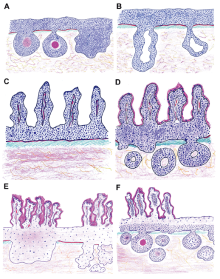
Schematic representations of the most frequent histologic subtypes of penile human papillomavirus-related squamous cell carcinomas. (A) Basaloid carcinoma mainly composed of small, blue cells grouped in solid sheets, or nests with central abrupt keratinization or comedo-like necrosis; (B) Basaloid carcinoma with small, blue cells grouped in solid sheets and trabeculae; (C) Papillary basaloid carcinoma—variant of the basaloid with papillae lined by small, blue cells; (D) Papillary basaloid carcinoma—abrupt keratinization and invasive nests can be seen; (E) Warty carcinoma—papillomatosis and infiltrative nests composed of koilocytes and cells with ample eosinophilic or clear cytoplasm are the hallmark; (F) Warty-basaloid carcinoma—features of warty and basaloid carcinomas are seen in the same specimen.
|

|
|
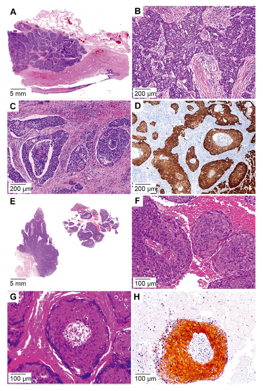
Basaloid squamous cell carcinoma and papillary basaloid variant. (A) Basaloid carcinoma seen as deeply invasive sheets, nests of interanastomosing trabeculae; (B) Starry night features seen due to this high-grade tumor; (C) Invasive nest with central comedo-like necrosis and characteristic surrounding clear space artifact; (D) p16 immunostain positive nests; (E) Low power view depicting the characteristic architecture of papillary basaloid carcinoma; (F) Papillae composed by a central fibrovascular core lined by small blue cells; (G) Abrupt keratinization and scant koilocytes; (H) p16 immunostain positive nest.
|
|
|
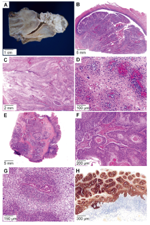
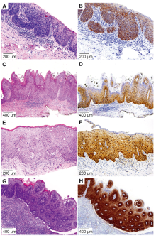
Warty and warty-basaloid squamous cell carcinoma human papillomavirus-related subtypes. (A) Papillomatous lesions arising from the foreskin; (B) Papillae seen at low power view; (C) The cells with ample eosinophilic to clear cytoplasm; (D) Atypical koilocytes surrounding a central fibrovascular core or arranged in invasive nests seen at higher power; (E) Warty-basaloid tumor with classic warty features on the surface and invasive basaloid nests in the lower left corner; (F) Both cell types (basaloid and warty) seen in the same sheet or nest; (G) Basaloid cells seen closer to the fibrovascular core and warty cells composing the superficial layers; (H) p16 immunostain of the basaloid component in warty-basaloid squamous cell carcinoma.
|

|
|
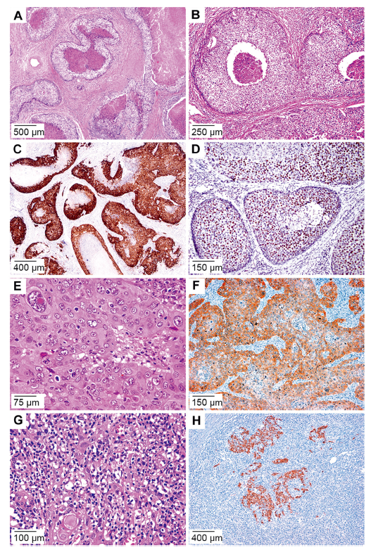
Less frequent HPV-related carcinomas histologic types. (A) Clear cell SCC with invasive solid sheets or nests; (B) Clear cell SCC nests composed of cells with ample clear cytoplasm and atypical koilocytes, abrupt keratinization and/or comedo-like necrosis; (C) p16 immunostain positive in the viable clear cells; (D) Chromogenic in situ hybridization for high-risk HPV in clear cell SCC; (E) Medullary carcinoma composed of solid nest of large, poorly differentiated cells and inflammatory cell infiltrate; (F) p16 immunostaining of the tumor cell sheets in medullary SCC; (G) Lymphoepithelioma-like carcinoma showing an inflammatory background with high grade tumor cells isolated or arranged in syncytial trabeculae; (H) p16 immunostain of squamous cells in the lymphoepithelioma-like carcinoma. HPV, human papillomavirus; SCC, squamous cell carcinoma.
|
| Subtype | Number | HPV genotype | Reference | | Basaloid | 104 | 16, 31, 33, 34, 35, 52, 55, 58, 73, 16+6, 16+33, 16+35, 16+56, 16+53, 16+44+52, 16+44+66, 18+52, 51+58 | -Alemany et al. [7], Fernández-Nestosa et al. [44] | | Warty | 33 | 6, 16, 33, 45, 52, 74, 35, 16+56, 31+33, 31+58, 59+74 | -Alemany et al. [7], Fernández-Nestosa et al. [44] | | Warty-basaloid | 26 | 16, 18, 35, 53, 59, 73, 16+70 | -Alemany et al. [7], Fernández-Nestosa et al. [44] | | Papillary basaloid | 11 | 16, 51, 16+45 | -Alemany et al. [7], Fernández-Nestosa et al. [44] | | Medullary | 12 | 16, 33, 58, 16+66 | -Ca?ete-Portillo et al. [8] | | Clear cell carcinoma | 8 | 16 | -Liegl and Regauer [19], Sanchez et al. [20] | | Lymphoepithelioma-like | 2 | High-risk HPV | -Mentrikoski et al. [9], | | Mixed | 33 | 16, 18, 26, 33, 39, 45, 52, 53, 58, 59 | Alemany et al. [7], Fernández-Nestosa et al. [44] |
|
|
HPV genotypes according to subtypes of invasive carcinoma.
|
|
|
Human papillomavirus-related subtypes of PeIN. (A) Basaloid PeIN. The same cellular features seen in the invasive counterpart were visible in hematoxylin and eosin stains; (B) p16 immunostain of the whole thickness of the epithelium in basaloid PeIN; (C) Warty-PeIN with papillary features; (D) p16 immunostain of basal cells in warty PeIN; (E) Warty PeIN with a flat surface. Note the clear cells corresponding to koilocytes without basaloid blue cells; (F) p16 immunostaining positive but weak of all cells in warty PeIN; (G) Warty-basaloid PeIN. Note the dual composition of this neoplasia, basaloid cells in the lower to mid part of the lesion and condylomatous cells in the surface; (H) p16 immunostaining strongly positive of the small basaloid cells in warty-basaloid PeIN. PeIN, penile intraepithelial neoplasias.
|
| Subtype | Number | HPV genotypesa | | Basaloid | 91 | 16, 18, 31, 33, 35, 44, 52, 56, 58, 16+54, 16+18+45+53, 16+51, 16+56, 16+53, 6+73, 16+18, 16+31, 16+53+56 | | Warty-basaloid | 49 | 16, 18, 30, 33, 35, 52, 56, 58, 51+52, 16+18+31, 31+51+53+58+66, 44+51+59 | | Warty | 29 | 16, 56, 39, 11, 18, 30, 33, 66, 73, 84, 87, 16+52+66, 18+73 | | Hybrid warty with basaloid | 2 | 16 | | Mixed | 1 | 16 |
|
|
HPV genotypes according to penile intraepithelial neoplasia subtypes.
|
| [1] |
Park JS, Jones RW, McLean MR, Currie JL, Woodruff JD, Shah KV, et al. Possible etiologic heterogeneity of vulvar intraepithelial neoplasia. A correlation of pathologic characteristics with human papillomavirus detection by in situ hybridization and polymerase chain reaction. Cancer 1991; 67: 1599e607.
|
| [2] |
Park JS, Rader JS, Wu TC, Laimins LA, Currie JL, Kurman RJ, et al. HPV-16 viral transcripts in vulvar neoplasia: preliminary studies. Gynecol Oncol 1991; 42:250e5.
|
| [3] |
Gregoire L, Cubilla AL, Reuter VE, Haas GP, Lancaster WD. Preferential association of human papillomavirus with highgrade histologic variants of penile-invasive squamous cell carcinoma. J Natl Cancer Inst 1995; 87:1705e9.
|
| [4] |
Rubin MA, Kleter B, Zhou M, Ayala G, Cubilla AL, Quint WGV, et al. Detection and typing of human papillomavirus DNA in penile carcinoma evidence for multiple independent pathways of penile carcinogenesis. Am J Pathol 2001; 159: 1211e8.
|
| [5] |
Cubilla AL, Lloveras B, Alejo M, Clavero O, Chaux A, Kasamatsu E, et al. The basaloid cell is the best tissue marker for human papillomavirus in invasive penile squamous cell carcinoma: a study of 202 cases from Paraguay. Am J Surg Pathol 2010; 34:104e14.
|
| [6] |
Olesen TB, Sand FL, Rasmussen CL, Albieri V, Toft BG, Norrild B, et al. Prevalence of human papillomavirus DNA and p16INK4a in penile cancer and penile intraepithelial neoplasia: a systematic review and meta-analysis. Lancet Oncol 2018; 20:145e58.
|
| [7] |
Alemany L, Cubilla A, Halec G, Kasamatsu E, Quirós B, Masferrer E, et al. Role of human papillomavirus in penile carcinomas worldwide. Eur Urol 2016; 69:953e61.
|
| [8] |
Ca?ete-Portillo S, Clavero O, Sanchez DF, Silvero A, Abed F, Rodriguez IM, et al. Medullary carcinoma of the penis: a distinctive HPV-related neoplasm:a report of 12 cases. Am J Surg Pathol 2017; 41:535e40.
|
| [9] |
Mentrikoski MJ, Frierson HF, Stelow EB, Cathro HP. Lymphoepithelioma-like carcinoma of the penis: association with human papilloma virus infection. Histopathology 2014; 64: 312e5.
|
| [10] |
Cubilla AL, Lloveras B, Alejo M, Clavero O, Chaux A, Kasamatsu E, et al. Value of p16INK4a in the pathology of invasive penile squamous cell carcinomas: a report of 202 cases. Am J Surg Pathol 2011; 35:253e61.
|
| [11] |
Canete-Portillo S, Velazquez EF, Kristiansen G, Egevad L, Grignon D, Chaux A, et al. Report from the International Society of Urological Pathology (ISUP) consultation conference on molecular pathology of urogenital cancers. Am J Surg Pathol 2020; 44:e80e6. https://doi.org/10.1097/PAS.0000000000001477.
|
| [12] |
Cubilla AL. Tumors of the penis. In: Epstein JI, Magi-Galluzzi C, Zhou M, Cubilla AL, editors. Tumors of the prostate gland, seminal vesicles, penis, and scrotum. Armed Forces Institute of Pathology (AFIP). 5th ed. Virginia: American Registry of Pathology; 2020. p. 405e612.
|
| [13] |
Cubilla AL, Reuter VE, Gregoire L, Ayala G, Ocampos S, Lancaster WD, et al. Basaloid squamous cell carcinoma: a distinctive human papilloma virus-related penile neoplasm. Am J Surg Pathol 1998; 22:755e61.
|
| [14] |
Alvarado-Cabrero I, Sanchez DF, Piedras D, Rodriguez-Gómez A, Rodriguez IM, Fernandez-Nestosa MJ, et al. The variable morphological spectrum of penile basaloid carcinomas:differential diagnosis, prognostic factors and outcome report in 27 cases classified as classic and mixed variants. Appl Cancer Res 2017; 37:3. https://doi.org/10.1186/s41241-017-0010-3.
|
| [15] |
Cubilla AL, Lloveras B, Alemany L, Alejo M, Vidal A, Kasamatsu E, et al. Basaloid squamous cell carcinoma of the penis with papillary features: a clinicopathologic study of 12 cases. Am J Surg Pathol 2012; 36:869e75.
|
| [16] |
Cubilla AL, Velazques EF, Reuter VE, Oliva E, Mihm MC, Young RH. Warty (condylomatous) squamous cell carcinoma of the penis:a report of 11 cases and proposed classification of “verruciform” penile tumors. Am J Surg Pathol 2000; 24: 505e12.
|
| [17] |
Manipadam MT, Bhagat SK, Gopalakrishnan G, Kekre NS, Chacko NK, Prasanna S. Warty carcinoma of the penis: a clinicopathological study from South India. Indian J Urol 2013; 29:282e7.
|
| [18] |
Chaux A, Tamboli P, Ayala A, Soares F, Rodriguez I, Barreto J, et al. Warty-basaloid carcinoma: clinicopathological features of a distinctive penile neoplasm. Report of 45 cases. Mod Pathol 2010; 23:896e904.
|
| [19] |
Liegl B, Regauer S. Penile clear cell carcinoma:a report of 5 cases of a distinct entity. Am J Surg Pathol 2004; 28:1513e7.
|
| [20] |
Sanchez DF, Rodriguez IM, Piris A, Ca?ete S, Lezcano C, Velazquez EF, et al. Clear cell carcinoma of the penis: an HPVrelated variant of squamous cell carcinoma. Am J Surg Pathol 2016; 40:917e22.
|
| [21] |
Schmauz R, Findlay M, Lalwak A, Katsumbira N, Buxton E. Variation in the appearance of giant condyloma in an Ugandan series of cases of carcinoma of the penis. Cancer 1977; 40: 1686e96.
|
| [22] |
Brennan S, Baird AM, O’Regan E, Sheils O. The role of human papilloma virus in dictating outcomes in head and neck squamous cell carcinoma. Front Mol Biosci 2021; 8:677900. https://doi.org/10.3389/fmolb.2021.677900.
|
| [23] |
Julia CJ, Hoang LN. A review of prognostic factors in squamous cell carcinoma of the vulva: evidence from the last decade. Semin Diagn Pathol 2020; 38:37e49.
|
| [24] |
Mohanty SK, Mishra SK, Bhardwaj N, Sardana R, Jaiswal S, Pattnaik N, et al. p53 and p16ink4a as predictive and prognostic biomarkers for Nodal metastasis and survival in a contemporary cohort of penile squamous cell carcinoma. Clin Genitourin Cancer 2021; 19:510e20.
|
| [25] |
Eich ML, Del Carmen Rodriguez Pena M, Schwartz L, Granada CP, Rais-Bahrami S, Giannico G, et al. Morphology, p16, HPV, and outcomes in squamous cell carcinoma of the penis: a multi-institutional study. Hum Pathol 2020; 96: 79e86.
|
| [26] |
Zargar-Shoshtari K, Spiess PE, Berglund AE, Sharma P, Powsang JM, Giuliano A, et al. Clinical significance of p53 and p16INK4a status in a contemporary North American penile carcinoma cohort. Clin Genitourin Cancer 2016; 14:346e51.
|
| [27] |
Zhang J, Zhang H, Xiu Y, Cheng H, Gu M, Song N. Prognostic significance of p16INK4a expression in penile squamous cell carcinoma: a meta-analysis with trial sequential analysis. Biomed Res Int 2018; 2018:8345893. https://doi.org/10.1155/2018/8345893.
|
| [28] |
Chu C, Chen K, Tan X, Lu J, Yang Y, Zhang Y, et al. Prevalence of human papillomavirus and implication on survival in Chinese penile cancer. Virchows Arch 2020; 477:667e75.
|
| [29] |
Sand FL, Rasmussen CL, Frederiksen MH, Andersen KK, Kj?r SK. Prognostic significance of HPV and p16 status in men diagnosed with penile cancer: a systematic review and metaanalysis. Cancer Epidemiol Biomarkers Prev 2018; 27: 1123e32.
|
| [30] |
Bezerra ALR, Lopes A, Santiago GH, Ribeiro KCB, Latorre MRDO, Villa LL. Human papillomavirus as a prognostic factor in carcinoma of the penis. Cancer 2001; 91:2315e21.
|
| [31] |
Bezerra SM, Chaux A, Ball MW, Faraj SF, Munari E, Gonzalez-Roibon N, et al. Human papillomavirus infection and immunohistochemical p16INK4a expression as predictors of outcome in penile squamous cell carcinomas. Hum Pathol 2015; 46: 532e40.
|
| [32] |
Steinestel J, Ghazal AA, Arndt A, Schnoeller TJ, Schrader AJ, Moeller P, et al. The role of histologic subtype, p16INK4a expression, and presence of human papillomavirus DNA in penile squamous cell carcinoma. BMC Cancer 2015; 15:220. https://doi.org/10.1186/s12885-015-1268-z.
doi: 10.1186/s12885-015-1268-z
pmid: 25885064
|
| [33] |
da Fonseca AG, Soares FA, Burbano RR, Silvestre RV, Pinto LOAD. Human papilloma virus: prevalence, distribution and predictive value to lymphatic metastasis in penile carcinoma. Int Braz J Urol 2013; 39:542e50.
|
| [34] |
Scheiner MA, Campos MM, Ornellas AA, Chin EW, Ornellas MH, Andrada-Serpa MJ. Human papillomavirus and penile cancers in Rio de Janeiro, Brazil: HPV typing and clinical features. Int Braz J Urol 2008; 34:467e76.
|
| [35] |
Guimaraes GC, Cunha IW, Soares FA, Lopes A, Torres J, Chaux A, et al. Penile squamous cell carcinoma clinicopathological features, nodal metastasis and outcome in 333 cases. J Urol 2009; 182:528e34.
|
| [36] |
Kravvas G, Ge L, Ng J, Shim TN, Doiron PR, Watchorn R, et al. The management of penile intraepithelial neoplasia (PeIN): clinical and histological features and treatment of 345 patients and a review of the literature. J Dermatolog Treat 2022; 33:1047e62.
|
| [37] |
Ashley S, Shanks JH, Oliveira P, Lucky M, Parnham A, Lau M, et al. Human papilloma virus (HPV) status may impact treatment outcomes in patients with precancerous penile lesions (an eUROGEN study). Int J Impot Res 2021; 33:620e6.
|
| [38] |
Soskin A, Vieillefond A, Carlotti A, Plantier F, Chaux A, Ayala G, et al. Warty/basaloid penile intraepithelial neoplasia is more prevalent than differentiated penile intraepithelial neoplasia in nonendemic regions for penile cancer when compared with endemic areas: a comparative study between pathologic series from Paris and Paraguay. Hum Pathol 2012; 43:190e6.
|
| [39] |
Chaux A, Velazquez EF, Amin A, Soskin A, Pfannl R, Rodriguez IM, et al. Distribution and characterization of subtypes of penile intraepithelial neoplasia and their association with invasive carcinomas: a pathological study of 139 lesions in 121 patients. Hum Pathol 2012 1; 43:1020e7.
|
| [40] |
Oertell J, Caballero C, Iglesias M, Chaux A, Amat L, Ayala E, et al. Differentiated precursor lesions and low-grade variants of squamous cell carcinomas are frequent findings in foreskins of patients from a region of high penile cancer incidence. Histopathology 2011; 58:925e33.
|
| [41] |
Singhal RR, Patel TM, Pariath KA, Vora RV. Premalignant male genital dermatoses. Indian J Sex Transm Dis AIDS 2019; 40: 97e104.
|
| [42] |
Velazquez EF, Soskin A, Bock A, Codas R, Barreto JE, Cubilla AL. Positive resection margins in partial penectomies: sites of involvement and proposal of local routes of spread of penile squamous cell carcinoma. Am J Surg Pathol 2004; 28: 384e9.
|
| [43] |
Chaux A, Reuter V, Lezcano C, Velazquez EF, Torres J, Cubilla AL. Comparison of morphologic features and outcome of resected recurrent and nonrecurrent squamous cell carcinoma of the penis: a study of 81 cases. Am J Surg Pathol 2009; 33:1299e306.
|
| [44] |
Fernández-Nestosa MJ, Guimerà N, Sanchez DF, Ca?ete-Portillo S, Velazquez EF, Jenkins D, et al. Human papillomavirus (HPV) genotypes in condylomas, intraepithelial neoplasia, and invasive carcinoma of the penis using laser capture microdissection (LCM)-PCR: a study of 191 lesions in 43 patients. Am J Surg Pathol 2017; 41:820e32.
|
| [45] |
Fernández-Nestosa MJ, Guimerà N, Sanchez DF, Ca?ete-Portillo S, Lobatti A, Velazquez EF, et al. Comparison of human papillomavirus genotypes in penile intraepithelial neoplasia and associated lesions: LCM-PCR study of 87 lesions in 8 patients. Int J Surg Pathol 2020; 28:265e72.
|
|
|
|