|
|
|
| The effect of perirenal fat stranding on infectious complications after ureterorenoscopy in patients with ureteral calculi |
Erhan Demirellia,*( ),Ercan Öğredena,Cemil Bayraktarb,Alptekin Tosunc,Ural Oğuza ),Ercan Öğredena,Cemil Bayraktarb,Alptekin Tosunc,Ural Oğuza
|
aGiresun University, Faculty of Medicine, Department of Urology, Giresun, Turkey
bMinistry of Health, Kayseri City Hospital, Department of Urology, Kayseri, Turkey
cGiresun University, Faculty of Medicine, Department of Radiology, Giresun, Turkey |
|
|
|
|
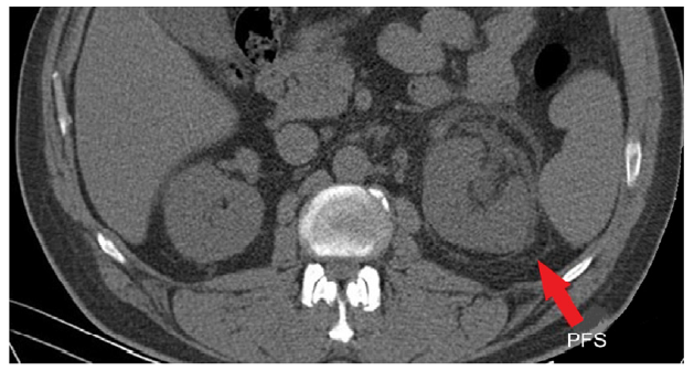
Abstract Objective: Perirenal fat stranding (PFS) is linear areas of soft-tissue attenuation in the perirenal space on non-contrast computed tomography. The present study aimed to investigate whether PFS is associated with infectious complications after ureterorenoscopy (URS) in patients with ureteral calculi in any location.
Methods: The data of 602 patients with ureteral stones who underwent URS were analyzed retrospectively. The patients were divided into two groups as Group 1 (PFS not detected) and Group 2 (PFS detected). Gender, and age of patients, size, side, and location of the stone, operation time, double-J stent insertion status, perioperative ureter injury, postoperative infection after URS and related complications, and duration of hospital stay were compared.
Results: While PFS was not detected in 530 patients, PFS was detected in 72 patients. The mean age, male/female ratio, side and localization of the stones, operation time, and perioperative insertion of the double-J after lithotripsy were statistically similar (p>0.05). The median stone diameter was smaller in Group 2 (9 mm vs. 8 mm) (p=0.033). Fever was observed in 30 and 38 patients in Group 1 and Group 2, respectively (p=0.0001). Urinary tract infection was detected in 24 and 27 patients in Group 1 and Group 2, respectively (p=0.0001). The urosepsis did not occur in any patients in Group 1, whereas 8 (11.1%) patients in Group 2 experienced urosepsis (p=0.0001).
Conclusion: According to the results of the present study, patients with ureteral stones accompanied by PFS are much more prone to ureteral injuries and infectious complications such as urinary tract infection, fever, and sepsis after URS.
|
|
Received: 20 February 2021
Available online: 20 July 2022
|
|
Corresponding Authors:
Erhan Demirelli
E-mail: erhandemirelli@yahoo.com
|
|
|

|
|
View of PFS in non-contrast computed tomography. PFS, perirenal fat stranding.
|
| Demographic information of patients and complications | Group 1 (n=530) | Group 2 (n=72) | p-Value | | Fever, n | | | 0.0001a | | Absent | 500 | 34 | | | Exist | 30 | 38 | | | Urinary infection, n | | | 0.0001a | | Absent | 506 | 45 | | | Exist | 24 | 27 | | | Urosepsis, n | | | 0.0001b | | Absent | 530 | 64 | | | Exist | 0 | 8 | | | Gender, n | | | 0.021a | | Female | 191 | 16 | | | Male | 339 | 56 | | | Stone side, n | | | 0.820a | | Left | 253 | 35 | | | Right | 275 | 37 | | | Bilateral | 2 | 0 | | | Stone localization, n | | | 0.145a | | Lower | 345 | 55 | | | Middle | 126 | 12 | | | Upper | 59 | 5 | | | Postoperative double-J stent insertion, n | | | 0.904a | | Absent | 375 | 52 | | | Exist | 155 | 20 | | | Mucosal injury, n | | | 0.0001b | | Absent | 517 | 54 | | | Exist | 13 | 18 | | | Ureteral perforation, n | | | 0.0001b | | Absent | 527 | 66 | | | Exist | 3 | 6 | | | Hydronephrosis, n | | | 0.076a | | Absent | 148 | 13 | | | Exist | 382 | 59 | | | Age, median, year | 44 | 43 | 0.772c | | Stone size, median, mm | 9 | 8 | 0.033c | | Operation time, median, min | 30 | 30 | 0.889c | | Postoperative length of hospital stay, median, day | 1 | 2 | 0.033c |
|
|
Patients’ data and postoperative infectious complications of two groups.
|
| Patients' data and risk factors | Complication | p-Value | | Absent | Exist | | Hydronephrosis, n | | | 0.688a | | Absent | 148 | 13 | | | Exist | 382 | 59 | | | PFS, n | | | 0.002a | | Absent | 467 | 63 | | | Exist | 53 | 19 | | | Gender, n | | | 0.045a | | Female | 187 | 20 | | | Male | 333 | 62 | | | Postoperative double-J stent insertion, n | | | 0.189a | | Absent | 374 | 53 | | | Exist | 146 | 29 | | | Age, mean, year | 44.9 | 44.2 | 0.685b | | Stone size, mean, mm | 8.8 | 10.4 | <0.001b | | Operation time, mean, min | 38.4 | 42.2 | 0.059b | | Interval time, mean, day | 14.4 | 14.9 | 0.867b |
|
|
Patients’ data and risk factors for postoperative infectious complications.
|
| Risk factor for infectious complication | Odds ratio | p-Value | 95% confidence interval | | Age, mean, year | 0.992 | 0.529 | 0.969-1.016 | | Gender | 1.032 | 0.930 | 0.516-2.064 | | Stone size, mean, mm | 1.089 | 0.041 | 1.003-1.182 | | Interval timea, mean, day | 1.002 | 0.910 | 0.969-1.036 | | Operation time, mean, min | 1.008 | 0.358 | 0.991-1.026 | | Postoperative double-J stent insertion | 2.765 | 0.013 | 1.245-6.143 | | PFS | 49.757 | <0.001 | 21.756-113.794 | | Presence of impacted stones | 7.765 | <0.001 | 3.146-19.167 |
|
|
Logistic regression analysis for all infectious complications (fever, urinary infection, and urosepsis).
|
| [1] |
Papadoukakis S, Stolzenburg J-U, Truss MC. Treatment strategies of ureteral stones. EAU-EBU Update Ser 2006; 4:184e90.
doi: 10.1016/j.eeus.2006.07.004
|
| [2] |
Nayyar R, Sarda AK, Kaza R, Anand V. The obstructed kidney. Indian J Surg 2005; 67:21e8.
|
| [3] |
Takahashi N, Kawashima A, Ernst RD, Boridy IC, Goldman SM, Benson GS, et al. Ureterolithiasis: can clinical outcome be predictedwith unenhanced helical CT? Radiology 1998; 208:97e102.
pmid: 9646798
|
| [4] |
Smith RC, Verga M, Dalrymple N, McCarthy S, Rosenfield AT. Acute ureteral obstruction: value of secondary signs of helical unenhanced CT. AJR Am J Roentgenol 1996; 167:1109e13.
doi: 10.2214/ajr.167.5.8911160
|
| [5] |
Yu TY, Kim HR, Hwang KE, Lee JM, Cho JH, Lee JH. Computed tomography findings associated with bacteremia in adult patients with a urinary tract infection. Eur J ClinMicrobiol Infect Dis 2016; 35: 1883e7.
|
| [6] |
Fukami H, Takeuchi Y, Kagaya S, Ojima Y, Saito A, Sato H, et al. Perirenal fat stranding is not a powerful diagnostic tool for acute pyelonephritis. Int J Gen Med 2017; 10:137e44.
doi: 10.2147/IJGM.S133685
|
| [7] |
Kadanalı A. [Üriner sistem infeksiyonları]. EAJM 2006; 38:119e23. [Article in Turkish].
|
| [8] |
Tepeler A, Resorlu B, Sahin T, Sarikaya S, Bayindir M, Oguz U, et al. Categorization of intraoperative ureteroscopy complications using modified Satava classification system. World J Urol 2014; 32:131e6.
doi: 10.1007/s00345-013-1054-y
|
| [9] |
TÜrk C, Pet_rk A, Sarica K, Seitz C, Skolarikos A, Straub M, et al. EAU guidelines on diagnosis and conservative management of urolithiasis. Eur Urol 2016; 69:468e74.
doi: 10.1016/j.eururo.2015.07.040
|
| [10] |
Hiller N, Berkovitz N, Lubashevsky N, Salaima S, Simanovsky N. The relationship between ureteral stone characteristics and secondary signs in renal colic. Clin Imag 2012; 36:768e72.
doi: 10.1016/j.clinimag.2012.01.018
|
| [11] |
Han NY, Sung DJ, Kim MJ, Park BJ, Sim KC, Cho SB. Perirenal fat stranding on CT: is there an association with bladder outlet obstruction? Br J Radiol 2016; 89:20160195. https://doi.org/10.1259/bjr.20160195.
|
| [12] |
Stunell H, Buckley O, Feeney J, Geoghegan T, Browne RF, Torreggiani WC. Imaging of acute pyelonephritis in the adult. Eur Radiol 2007; 17: 1820e8.
|
| [13] |
Kim JS, Lee S, Lee KW, Kim JM, Kim YH, Kim ME. Relationship between uncommon computed tomography findings and clinical aspects in patients with acute pyelonephritis. Korean J Urol 2014; 55:482e6.
doi: 10.4111/kju.2014.55.7.482
|
| [14] |
Hernandez N, Mozafarpour S, Song Y, Eisner BH. Cessation of ureteral colic does not necessarily mean that a ureteral stone has been expelled. J Urol 2018; 199:1011e4.
doi: S0022-5347(17)77794-4
pmid: 29107030
|
| [15] |
ö _greden E, O_guz U, Demirelli E, Benli E, Sancak EB, GÜlpinar MT, et al. Categorization of ureteroscopy complications and investigation of associated factors by using the modified Clavien classification system. Turk J Med Sci 2016; 46: 686e94.
|
| [16] |
Cindolo L, Castellan P, Scoffone CM, Cracco CM, Celia A, Paccaduscio A, et al. Mortality and flexible ureteroscopy: analysis of six cases. World J Urol 2016; 34:305e10.
doi: 10.1007/s00345-015-1642-0
pmid: 26210344
|
| [17] |
Cindolo L, Castellan P, Primiceri G, Hoznek A, Cracco CM, Scoffone CM, et al. Life-threatening complications after ureteroscopy for urinary stones: survey and systematic literature review. Minerva Urol Nefrol 2017; 69:421e31.
doi: 10.23736/S0393-2249.17.02787-4
pmid: 28150482
|
| [18] |
Berardinelli F, De Francesco P, Marchioni M, Cera N, Proietti S, Hennessey D, et al. Infective complications after retrograde intrarenal surgery: a new standardized classification system. Int Urol Nephrol 2016; 48:1757e62.
pmid: 27443315
|
| [19] |
Mandal S, Goel A, Singh MK, Kathpalia R, Nagathan DS, Sankhwar SN, et al. Clavien classification of semirigid ureteroscopy complications: a prospective study. Urology 2012; 80:995e1001.
doi: 10.1016/j.urology.2012.05.047
|
| [20] |
Fan S, Gong B, Hao Z, Zhang L, Zhou J, Zhang Y, et al. Risk factors of infectious complications following flexible ureteroscope with a holmium laser: a retrospective study. Int J Clin Exp Med 2015; 8:11252e9.
|
| [21] |
Martov A, Gravas S, Etemadian M, Unsal A, Barusso G, D’Addessi A, et al. Postoperative infection rates in patients with a negative baseline urine culture undergoing ureteroscopic stone removal: a matched case-control analysis on antibiotic prophylaxis from the CROES URS global study. J Endourol 2015; 29:171e80.
doi: 10.1089/end.2014.0470
|
| [22] |
de la Rosette J, Denstedt J, Geavlete P, Keeley F, Matsuda T, Pearle M, et al. The clinical research office of the endourological society ureteroscopy global study: indications, complications, and outcomes in 11 885 patients. J Endourol 2014; 28:131e9.
doi: 10.1089/end.2013.0436
pmid: 24147820
|
| [23] |
Southern JB, Higgins AM, Young AJ, Kost KA, Schreiter BR, Clifton M, et al. Risk factors for postoperative fever and systemic inflammatory response syndrome after ureteroscopy for stone disease. J Endourol 2019; 33:516e22.
doi: 10.1089/end.2018.0789
pmid: 30569755
|
| [24] |
Beysens M, Tailly TO. Ureteral stents in urolithiasis. Asian J Urol 2018; 5:274e86.
|
| [25] |
Ogreden E, Oguz U, Demirelli E, Benli E, ö zen ö. The impact of ureteral double-J stent insertion following ureterorenoscopy in patients with ureteral stones accompanied by perirenal fat stranding. Arch Ital Urol Androl 2018; 90:15e9.
doi: 10.4081/aiua.2018.1.15
|
| [26] |
Nevo A, Mano R, Baniel J, Lifshitz DA. Ureteric stent dwelling time: a risk factor for post-ureteroscopy sepsis. BJU Int 2017; 120:117e22.
doi: 10.1111/bju.13796
|
| [27] |
Rashid AO, Fakhulddin SS. Risk factors for fever and sepsis after percutaneous nephrolithotomy. Asian J Urol 2016; 3:82e7.
doi: 10.1016/j.ajur.2016.03.001
pmid: 29264169
|
|
|
|