a Department of Urology, King George's Medical University, Lucknow, Uttar Pradesh, India b Department of Nuclear Medicine, Dr. Ram Manohar Lohia Institute of Medical Sciences, Lucknow, India c Department of Obstetrics and Gynaecology, King George's Medical University, Lucknow, Uttar Pradesh, India
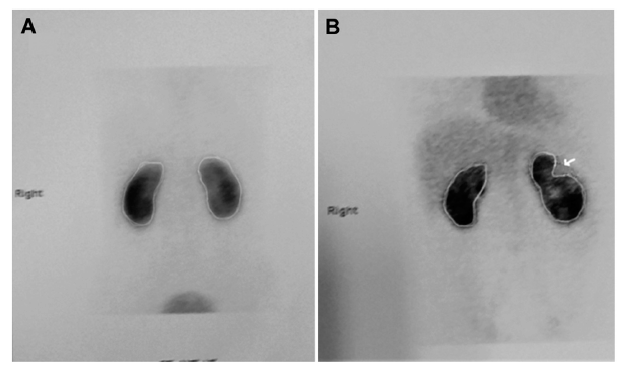
Objective: To look for change in relative renal function and document renal scarring following endoscopic renal pelvic instillation sclerotherapy (RPIS) in patients with chyluria by dimercaptosuccinic acid (DMSA) renal scan. Methods: A prospective study was performed between November 2015 and September 2016. All patients with biochemically documented chyluria who underwent RPIS using either 1%-silver nitrate or 0.1%-povidine iodine were included. Patients received either 3-, 6- or 9-doses. DMSA renal scan was performed before and 2-3 months after sclerotherapy. Results: Of the 34 patients, 22 were males. Mean age was 41.08 ± 16.64 years (range, 15-70 years). Thirty-two patients (94.1%) responded to therapy while two did not respond even after 9-doses. Average follow-up was 8.94 ± 3.70 months. The mean relative renal function (pre-instillation) of normal kidney was 50.76% ± 3.55% while that of affected renal unit (side of instillation) was 49.20% ± 3.44% (range, 43.0%-61.0%). After instillation therapy, the mean relative renal function of normal side was 52.26% ± 3.57% while that of affected renal unit was 47.50% ± 3.56% (range, 41.0%-54.0%). The relative renal function did not change >5% from the baseline value in any patient except one (in which the differential function increased paradoxically by 12%). Two patients developed renal scar in post-instillation renal scan. Conclusion: Endoscopic sclerotherapy in chyluria is safe and effective. The relative renal function does not deteriorate by more than 5%. There is a small risk of development of renal scar. More studies involving larger number of patients are needed to answer this dilemma.
. [J]. Asian Journal of Urology, 2019, 6(4): 359-363.
Bimalesh Purkait,Apul Goel,Satyawati Deswal,Monica Agrawal,BhupendraPal Singh,Manoj Kumar. Does endoscopic sclerotherapy in filarial chyluria affect renal function and morphology? A prospective study using dimercaptosuccinic acid renal scan. Asian Journal of Urology, 2019, 6(4): 359-363.
Ngan H, Yu HHY, Leong CH . A lymphographic study of chyluria. Br J Radiol 1977; 50:863-70.
[3]
Sharma G, Chitale V, Karva R, Sharma A, Durug AB . Fluoroscopy guided instillation therapy in chyluria using combination of povidone iodine with contrast agent. Is a single instillation sufficient? Int Braz J Urol 2008; 34:270-5.
[4]
Sabnis RB, Punekar SV, Desai RM, Bradoo AM, Bapat SD . Instillation of silver nitrate in the treatment of chyluria. Br J Urol 1992; 70:660-2.
[5]
Shanmugam TV, Prakash JV, Sivashankar G . Povidone iodine used as a sclerosing agent in the treatment of chyluria. Br J Urol 1998; 82:587.
[6]
Goel S, Mandhani A, Srivastava A, Kapoor R, Gogoi S, Kumar A , et al. Is povidone iodine an alternative to silver nitrate for renal pelvic instillation sclerotherapy in chyluria? BJU Int 2004; 94:1082-5.
[7]
Dash SC, Bhargav Y, Saxena S, Agarwal SK, Tiwari SC, Dinda A . Acuterenal failure and renal papillary necrosis following instillation of silver nitrate for treatment of chyluria. Nephrol Dial Transplant 1996; 11:1841-2.
[8]
Kulkarni AA, Pathak MS, Sirsat RA . Fatal renal and hepatic failure following silver nitrate instillation for treatment of chyluria. Nephrol Dial Transplant 2005; 20:1276-7.
[9]
Kumar M, Bhat HS . Experience with 5% AgNO3 instillation in chyluria. Indian J Surg 1975; 37:33-8.
[10]
Mandhani A, Kapoor R, Gupta RK, Rao HS . Can silver nitrate instillation for the treatment of chyluria be fatal? Br J Urol 1998; 82:926-7.
[11]
Moorin R . 99mTc-DMSA absolute uptake: normal pediatric values at 24 hours. J Nucl Med Technol 2001; 29:22-9.
[12]
Nepple KG, Knudson MJ, Austin JC, Cooper CS . Abnormal renal scans and decreased early resolution of low grade vesicoureteral reflux. J Urol 2008; 180:1643-7.
[13]
Goel TC, Goel A. Chyluria. In: Goel TC, Goel A, editors. Lymphatic filariasis. Springer: Singapore; 2016. p. 273-300.
[14]
Desai R . Complications and precautions of sclerotherapy. Indian J Urol 2005; 21:27-30.
[15]
Shanon A, Feldman W, McDonald P, Martin DJ, Matzinger MA, Shillinger JF , et al. Evaluation of renal scars by technetiumlabeled dimercapto-succinic acid scan, intravenous urography, and ultrasonography: a comparative study. J Pediatr 1992; 120:399-403.
[16]
Rossleigh MA . Renal cortical scintigraphy and diuresis renography in infants and children. J Nucl Med 2001; 42:91-5.
[17]
Bhatnagar V, Mitra DK, Agarwala S, Kumar R, Patel C, Malhotra AK , et al. The role of DMSA scans in evaluation of the correlation between urinary tract infection, vesicoureteric reflux, and renal scarring. Pediatr Surg Int 2002; 18:128-34.
[18]
Matsura H, Hioki T, Sakurai M, Arima K, Yanagawa M, Sugimura Y , et al. Influence of extracorporeal shock wave lithotripsy (ESWL) on renal function assessed by 99mTc-DMSA scintigraphy: comparative analysis between ESWL and percutaneous nephroureterolithotripsy (PNL). Hinuokyka Kiyo 1994; 40:1061-7.
[19]
Mousavi-bahar SH, Fazlian MM . Effect of percutaneous nephrolithotomy on renal function measured by Tc-99m-Dimercapto- Succinic cid renal scan. Internet J Nephrol 2010; 2:234-8.
[20]
Jacobson SH, Hansson S, Jakobsson B . Vesicoureteric reflux: occurrence and long term risks. Acta Paediatr Suppl 1999; 43:22-30.
[21]
Zaki M, Badawi M, Al Mutari G, Ramadan D, Adul Rahman M . Acute pyelonephritis and renal scarring in Kuwaiti children: a follow-up study using 99mTc DMSA renal scintigraphy. Pediatr Nephrol 2005; 20:1116-9.
[22]
Stokland E, Hellstróm M, Jacobson B . Early Tc-99m DMSA scintigraphy in symptomatic first-time urinary tract infection. Acta Paediatr 1996; 85:430-6.
[23]
Stokland E, Hellstro′m M, Jacobson B . Renal damage one year after first urinary tract infection, role of DMSA scintigraphy. J Pediatr 1996; 129:815-20.
[24]
Camacho V, Estorch M, Fraga G, Mena E, Fuertes J, Hernández MA , et al. DMSA study performed during febrile urinary tract infection:a predictor of patient outcome? Eur J Nucl Med Mol Imaging 2004; 31:862-6.