|
|
|
| Prospective observational study on the prognosis of ureteral lesions caused by impacted stones via dual-energy spectral computed tomography |
Junjie Wanga,Ximing Wangb,Haozhou Zhongc,Wengui Xiec,Qilin Xic,*( ) )
|
aDepartment of Urology, The Sixth People's Hospital of Chengdu, Chengdu, China
bDepartment of Radiology, The First Affiliated Hospital of Soochow University, Suzhou, China
cDepartment of Urology, The First Affiliated Hospital of Soochow University, Suzhou, China |
|
|
|
|
Abstract Objective: Ureteral lesions caused by impacted ureteral stones are likely to result in postoperative ureteral stricture. On this basis, the study aimed to investigate if dual-energy spectral computed tomography can predict ureteral hardening caused by impacted stones and to explore the relationship between different types of ureteral lesions and the risk of ureteral stricture. Methods: This prospective study collected data of 93 patients with impacted stones from hospital automation system during January 2018 to October 2019. They underwent an abdominal scan on a dual-energy spectral computed tomography. During surgery, the operator used ureteroscopy to identify ureteral lesions, which were classified into four categories: edema, polyps, pallor, and hardening. Seven months later, 90 patients were reviewed for the degree of hydronephrosis. Results: Endoscopic observations revealed 38 (41%) cases of ureteral edema, 20 (22%) cases of polyps, 13 (14%) cases of pallor, and 22 (24%) cases of hardening. There were significant differences in hydronephrosis, the period of impaction, the calcium concentration of the ureter, and the slope of the spectral Hounsfield unit curve between the four groups. After that, we evaluated the factors associated with ureteral hardening and found that the calcium concentration of the ureter and hydronephrosis remained independent predictors of ureteral hardening. Receiver operating characteristic curve analysis showed that 5.3 mg/cm³ calcium concentration of the ureter is an optimal cut-off value to predict ureteral hardening. The result of follow-up showed that 80 patients had complete remission of hydronephrosis, with a complete remission rate of 61.9% (13/21) in the hardening group and 97.1% (67/69) in the non-hardening group (p<0.001). Conclusion: Calcium concentration of the ureter is an independent predictor of ureteral hardening. Patients with ureteral hardening have more severe hydronephrosis after ureteroscopic lithotripsy. When the calcium concentration of the ureter is less than 5.3 mg/cm³, ureteral lesions should be actively treated.
|
|
Received: 03 June 2021
Available online: 20 October 2023
|
|
Corresponding Authors:
*E-mail address: linqix_827@163.com (Q. Xi).
|
|
|
|
|
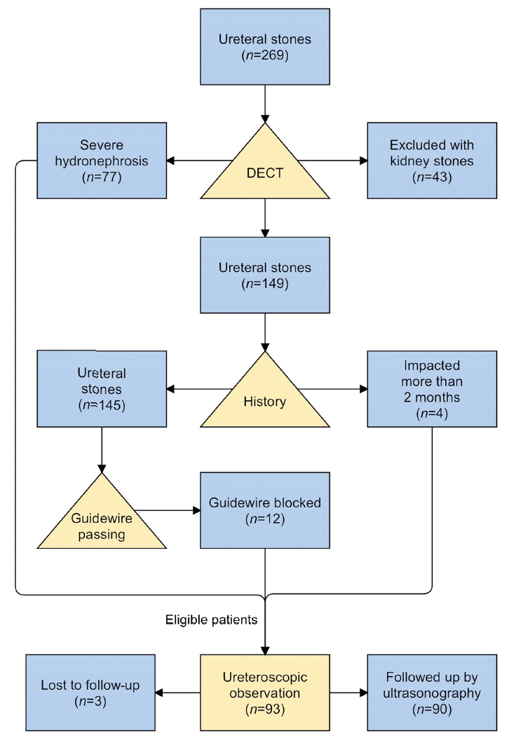
Participant flow. DECT, dual-energy spectral computed tomography.
|
|
|
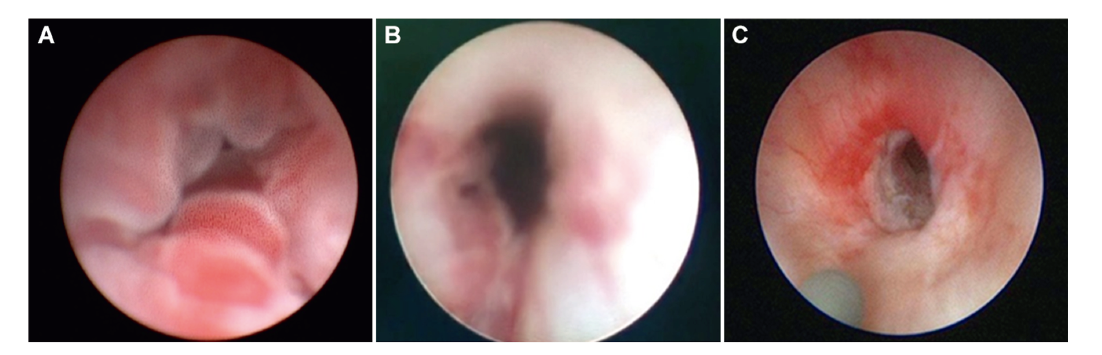
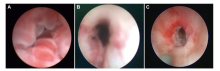
Endoscopic view of different ureteral lesions caused by impacted stones. (A) Villous or multiple stripy protrusions; (B) Pale and usually free of polyps; (C) Narrowed, stiff, and appeared interstitial fibrosis.
|
| Characteristic | Hardening | Pallor | Polyps | Edema | p-Value | | Patient, n | 22 | 13 | 20 | 38 | NA | | Mean age, year | 56 | 58 | 48 | 50 | 0.105 | | Sex, n | | | | | 0.373 | | Female | 11 | 4 | 6 | 11 | | | Male | 11 | 9 | 14 | 27 | | | Site of impaction, n | | | | | 0.06 | | Lower ureter | 1 | 1 | 6 | 14 | | | Middle ureter | 5 | 2 | 1 | 5 | | | Upper ureter | 16 | 10 | 13 | 19 | | | Stone sizea,b, mm | 13.6±3.7 | 13.7±5.9 | 13.0±5.9 | 11.7±4.4 | 0.178 | | Hydronephrosisa, mm | 38.9±15.1 | 36.4±14.3 | 28.3±8.9 | 24.5±8.5 | <0.001c | | Duration of symptomsa, month | 3.4±5.8 | 4.6±5.0 | 2.9±4.3 | 1.3±2.4 | 0.037c | | Stone diametera, mm | 9.6±2.3 | 9.9±3.8 | 9.4±3.2 | 8.1±3.1 | 0.152 | | Ureteral wall thicknessa, mm | 8.1±3.2 | 8.8±3.1 | 9.5±3.6 | 8.1±2.4 | 0.629 | | Spectral CT | | | | | | | Calcium concentrationa, mg/cm3 | 3.7±3.4 | 6.1±2.8 | 8.5±2.7 | 8.2±3.1 | <0.001c | | Water concentrationa, mg/cm3 | 1007.3±21.6 | 1009.6±11.5 | 1019.0±15.0 | 1013.3±13.8 | 0.214 | | Effective-Za | 7.8±0.3 | 7.9±0.1 | 8.0±0.2 | 8.0±0.3 | 0.114 | | λHUa | 0.8±0.6 | 1.0±0.6 | 1.4±0.6 | 1.4±0.5 | 0.038c |
|
|
Baseline characteristics of hardening, pallor, polyps, and edema groups.
|
| Variablea | p-Value | | Hydronephrosis | | Hardening vs. edema | <0.001b | | Pallor vs. edema | 0.018b | | Calcium concentration | | Hardening vs. edema | <0.001b | | Hardening vs. polyps | <0.001b | | Rate of curve | | Hardening vs. polyps | 0.038b | | Hardening vs. edema | 0.017b | | Duration of impaction | | Pallor vs. edema | 0.037b |
|
|
Results from pairwise comparisons between groups.
|
| Variable | OR (95% CI) | p-Value | | Hydronephrosis, cm | 1.063 (1.012-1.115) | 0.014a | | Duration of symptoms, month | 1.088 (0.923-1.282) | 0.314 | | Calcium concentration, mg/cm3 | 0.595 (0.432-0.820) | 0.001a | | λHU | 1.642 (0.364-7.414) | 0.378 |
|
|
Multivariate logistic regression analysis of preoperative factors associated with the hardening and non-hardening.
|
|
|
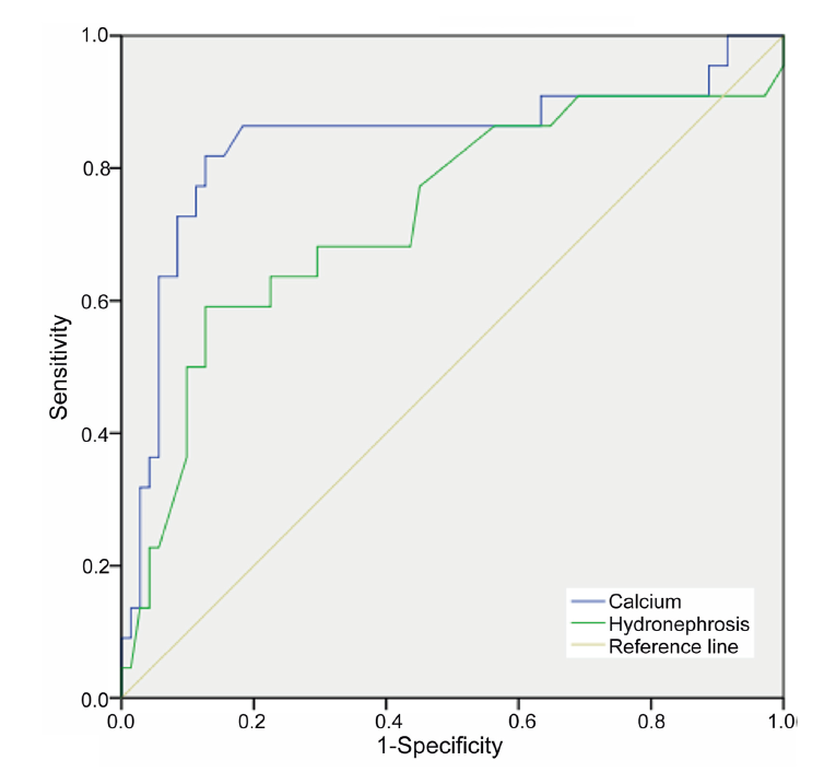
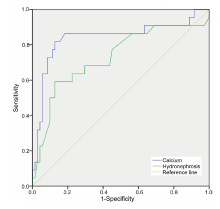
Effect of dual-energy spectral computed tomography calcium concentration of the ureteral wall and hydronephrosis on preoperative prediction of ureteral hardening.
|
| Group | Calcium concentration | | <5.3 mg/cm3 | ≥5.3 mg/cm3 | | Hardening, n | 18 | 4 | | Pallor, n | 4 | 9 | | Polyps, n | 2 | 18 | | Edema, n | 3 | 35 |
|
|
Distribution of calcium concentration of dual-energy spectral computed tomography in four groups.
|
| [1] |
Sorokin I, Mamoulakis C, Miyazawa K, Rodgers A, Talati J, Lotan Y. Epidemiology of stone disease across the world. World J Urol 2017; 35:1301-20.
doi: 10.1007/s00345-017-2008-6
pmid: 28213860
|
| [2] |
Wang W, Fan J, Huang G, Li J, Zhu X, Tian Y, et al. Prevalence of kidney stones in mainland China: a systematic review. Sci Rep 2017; 7:41630.
doi: 10.1038/srep41630
pmid: 28139722
|
| [3] |
Legemate JD, Wijnstok NJ, Matsuda T, Strijbos W, Erdogru T, Roth B, et al. Characteristics and outcomes of ureteroscopic treatment in 2650 patients with impacted ureteral stones. World J Urol 2017; 35:1497-506.
doi: 10.1007/s00345-017-2028-2
pmid: 28321499
|
| [4] |
Darwish AE, Gadelmoula MM, Abdelkawi IF, Abdellatif AM, Abdel-Moneim AM, Hammouda HM. Ureteral stricture after ureteroscopy for stones: a prospective study for the incidence and risk factors. Urol Ann 2019; 11:276-81.
doi: 10.4103/UA.UA_110_18
pmid: 31413506
|
| [5] |
?zbir S, Can O, Atalay HA, Canat HL, ?ak?r SS, ?tün?temur A. Formula for predicting the impaction of ureteral stones. Urolithiasis 2020;48:353-60.
|
| [6] |
Mugiya S, Ito T, Maruyama S, Hadano S, Nagae H. Endoscopic features of impacted ureteral stones. J Urol 2004; 171:89-91.
doi: 10.1097/01.ju.0000100960.08768.81
pmid: 14665851
|
| [7] |
Tas S, Tugcu V, Mutlu B, Karadag S, Bitkin A, Yucel M, et al. Incidence of ureteral stricture after ureterorenoscopic pneumatic lithotripsy for distal ureteral calculi. Arch Ital Urol Androl 2011; 83:141-6.
|
| [8] |
El-Abd AS, Suliman MG, Abo Farha MO, Ramadan AR, El-Tatawy HH, El-Gamal OM, et al. The development of ureteric strictures after ureteroscopic treatment for ureteric calculi: a long-term study at two academic centres. Arab J Urol 2014; 12: 168-72.
doi: 10.1016/j.aju.2013.11.004
|
| [9] |
Brito AH, Mitre AI, Srougi M. Ureteroscopic pneumatic lithotripsy of impacted ureteral calculi. Int Braz J Urol 2006; 32:295-9.
pmid: 16813672
|
| [10] |
Aldoukhi AH, Ghani KR, Hall TL, Roberts WW. Thermal response to high-power holmium laser lithotripsy. J Endourol 2017; 31:1308-12.
doi: 10.1089/end.2017.0679
pmid: 29048216
|
| [11] |
Machida H, Tanaka I, Fukui R, Shen Y, Ishikawa T, Tate E, et al. Dual-energy spectral CT: various clinical vascular applications. Radiographics 2016; 36:1215-32.
doi: 10.1148/rg.2016150185
pmid: 27399244
|
| [12] |
McCollough CH, Leng S, Yu L, Fletcher JG. Dual- and multienergy CT: principles, technical approaches, and clinical applications. Radiology 2015; 276:637-53.
doi: 10.1148/radiol.2015142631
pmid: 26302388
|
| [13] |
Fam XI, Singam P, Ho CC, Sridharan R, Hod R, Bahadzor B, et al. Ureteral stricture formation after ureteroscope treatment of impacted calculi: a prospective study. Korean J Urol 2015; 56:63-7.
doi: 10.4111/kju.2015.56.1.63
pmid: 25598938
|
| [14] |
Tran TY, Bamberger JN, Blum KA, Parkhomenko E, Thai J, Chandhoke RA, et al. Predicting the impacted ureteral stone with computed tomography. Urology 2019; 130:43-7.
doi: S0090-4295(19)30377-2
pmid: 31029671
|
| [15] |
Deguchi R, Yamashita S, Kikkawa K, Kohjimoto Y, Hara I. HU above-below ratio is an useful preoperative factor for predicting impacted ureteral calculi. Eur Urol Open Sci 2020; 19: e840-1. https://doi.org/10.1016/s2666-1683(20)33145-1.
|
| [16] |
May PC, Hsi RS, Tran H, Stoller ML, Chew BH, Chi T, et al. The morbidity of ureteral strictures in patients with prior ureteroscopic stone surgery: multi-institutional outcomes. J Endourol 2018; 32:309-14.
doi: 10.1089/end.2017.0657
pmid: 29325445
|
| [17] |
Sugino T, Taguchi K, Hamamoto S, Okada T, Isogai M, Tanaka Y, et al. Risk factors for failure of endoscopic management of stone-related ureteral strictures. Urol J 2021; 19:95-100.
doi: 10.1016/S0022-5347(17)73208-9
|
| [18] |
Elashry OM, Elgamasy AK, Sabaa MA, Abo-Elenien M, Omar MA, Eltatawy HH, et al. Ureteroscopic management of lower ureteric calculi: a 15-year single-centre experience. BJU Int 2008; 102:1010-7.
doi: 10.1111/j.1464-410X.2008.07747.x
pmid: 18485033
|
| [19] |
Al-Nabulsi Z, Phan YC, Abdalla O, Austin T, Tanasescu G, Osborn P, et al. Surgical and radiological predictive factors for ureteric stricture formation in patients treated with ureteroscopy for ureteric stones. Scand J Urol 2021; 55: 394-8.
|
| [20] |
Xi Q, Wang S, Ye Z, Liu J. Combined removal of stones with resection of concurrent pathologic ureter may be a preferred treatment for impacted ureteral stones with stricture lesions. J Endourol 2009; 23:243-7.
doi: 10.1089/end.2008.0507
pmid: 19220083
|
| [21] |
Chandhoke R, Bamberger JN, Gallante B, Atallah W, Gupta M. Peri-calculus ureteral thickness on computed tomography predicts stone impaction at time of surgery: a prospective study. J Endourol 2020; 34:107-11.
doi: 10.1089/end.2019.0449
pmid: 31650853
|
| [22] |
Selvi I, Baydilli N, Tokmak TT, Akinsal EC, Basar H. CT-related parameters and Framingham score as predictors of spontaneous passage of ureteral stones _10 mm: results from a prospective, observational, multicenter study. Urolithiasis 2021; 49:227-37.
doi: 10.1007/s00240-020-01214-6
|
| [23] |
Gong B, Wu Y, O’Keeffe ME, Berger FH, McLaughlin PD, Nicolaou S, et al. Top 50 highly cited articles on dual energy computed tomography (DECT) in abdominal radiology: a bibliometric analysis. Pol J Radiol 2017; 82:748-59.
doi: 10.12659/PJR.904075
|
| [24] |
Dretler SP, Young RH. Stone granuloma: a cause of ureteral stricture. J Urol 1993; 150:1800-2.
doi: 10.1016/s0022-5347(17)35899-8
pmid: 8230508
|
| [25] |
Morgentaler A, Bridge SS, Dretler SP. Management of the impacted ureteral calculus. J Urol 1990; 143:263-6.
doi: 10.1016/s0022-5347(17)39928-7
pmid: 1967657
|
| [26] |
Huffiman J. Abnormal ureter and intrarenal collecting system. Urol Endosc Manual Atlas 1985:59-73.
|
| [27] |
Shen Y, Xiang A, Shao S. Preoperative hydronephrosis is a predictive factor of ureteral stenosis after flexible ureteroscopy: a propensity scores matching analysis. BMC Urol 2021; 21:153. https://doi.org/10.1186/s12894-021-00917-1.
doi: 10.1186/s12894-021-00917-1
pmid: 34763687
|
| [28] |
Abat D, B?reko?lu A, Altunkol A, K?se I, Bo?a MS. Is there any predictive value of the ratio of the upper to the lower diameter of the ureter for ureteral stone impaction? Curr Urol 2021;15: 161-6.
|
| [29] |
Roberts WW, Cadeddu JA, Micali S, Kavoussi LR, Moore RG. Ureteral stricture formation after removal of impacted calculi. J Urol 1998; 159:723-6.
pmid: 9474134
|
| [30] |
Devarajan R, Ashraf M, Beck RO, Lemberger RJ, Taylor MC. Holmium:YAG lasertripsy for ureteric calculi: an experience of 300 procedures. Br J Urol 1998; 82:342-7.
pmid: 9772868
|
| [31] |
Yoshida T, Inoue T, Omura N, Okada S, Hamamoto S, Kinoshita H, et al. Ureteral wall thickness as a preoperative indicator of impacted stones in patients with ureteral stones undergoing ureteroscopic lithotripsy. Urology 2017; 106:45-9.
doi: S0090-4295(17)30471-5
pmid: 28499762
|
| [32] |
Weizer AZ, Auge BK, Silverstein AD, Delvecchio FC, Brizuela RM, Dahm P, et al. Routine postoperative imaging is important after ureteroscopic stone manipulation. J Urol 2002; 168:46-50.
pmid: 12050490
|
| [1] |
Gild Philipp,A. Kluth Luis,W. Vetterlein Malte,Engel Oliver,K.H. Chun Felix,Fisch Margit. Adult iatrogenic ureteral injury and stricture-incidence and treatment strategies[J]. Asian Journal of Urology, 2018, 5(2): 101-106. |
| [2] |
Dong Hao,Peng Yonghan,Li Ling,Gao Xiaofeng. Prevention strategies for ureteral stricture following ureteroscopic lithotripsy[J]. Asian Journal of Urology, 2018, 5(2): 94-100. |
| [3] |
Brian D. Duty, John M. Barry. Diagnosis and management of ureteral complications following renal transplantation[J]. Asian Journal of Urology, 2015, 2(4): 202-207. |
|
|
|
|