|
|
|
| Perinephric myxoid pseudotumor of fat in a young patient with a horseshoe kidney complicated by an obstructing renal calculus |
Ren Wei Liua,*( ),Gideon Tanb,Yi Ting Lima,Wei Jin Chuac,Amanda Chenga,Ying Mei Wonga,Victor Leeb,Thomas Thamboob ),Gideon Tanb,Yi Ting Lima,Wei Jin Chuac,Amanda Chenga,Ying Mei Wonga,Victor Leeb,Thomas Thamboob
|
a Department of Diagnostic Imaging, National University Hospital, Singapore
b Department of Pathology, National University Hospital, Singapore
c Department of Urology, National University Hospital, Singapore |
|
|
|
|
|
|
Received: 20 September 2021
Available online:
|
|
Corresponding Authors:
Ren Wei Liu
E-mail: REN_WEI_LIU@NUHS.EDU.SG
|
|
|
|
|
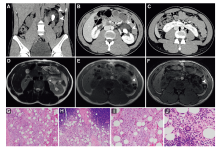
CT and magnetic resonance imaging of the abdomen and pelvis with intravenous contrast, and pathology images showing microscopic features of the lesion. (A and B) Coronal and axial CT images showing a large obstructing calculus in the left renal pelvis (black arrows) and a low-density perirenal soft tissue mass with enhancing components (white arrowheads); (C) Horseshoe configuration of the kidneys (black arrows); (D) T2-weighted axial magnetic resonance imaging showing vascular flow voids suggestive of vessels (white arrowheads); (E) In-phase and (F) out-of-phase magnetic resonance imaging sequences showing signal drop-out consistent with macroscopic fat content; (G-I) Pathological analysis showing mature adipose tissue with areas of myxoid change, small blood vessels, and aggregates of chronic inflammatory cells, and there was no significant stromal atypia; (J) Scattered Touton-type giant cells present in the lesion, predominantly in the areas with chronic inflammation. CT, computed tomography.
|
| [1] |
Mitreski G, Sutherland T.Radiological diagnosis of perinephric pathology: pictorial essay 2015. Insights Imaging 2017; 8: 155-69.
doi: 10.1007/s13244-016-0536-z
|
| [2] |
Israel GM, Bosniak MA. How I do it: evaluating renal masses. Radiology 2005; 236:441-50.
doi: 10.1148/radiol.2362040218
pmid: 16040900
|
| [3] |
Chen F, Desai MA, Cernigliaro JG, Edgar MA, Alexander LF. Perinephric myxoid pseudotumor of fat: a very rare entity that can mimic a renal cyst and retroperitoneal liposarcoma on imaging. Clin Imag 2021; 69:139-44.
doi: 10.1016/j.clinimag.2020.06.041
pmid: 32731105
|
| [4] |
Dashti NK, Fritchie KJ, Folpe AL. Perinephric myxoid pseudotumor of fat: a distinctive pseudoneoplasm most often associated with non-neoplastic renal disease. Hum Pathol 2019; 87: 37-43.
doi: S0046-8177(19)30022-X
pmid: 30826321
|
| [5] |
Tanas MR, Sthapanachai C, Nonaka D, Melamed J, Oliveira AM, Erickson-Johnson MR, et al. Pseudosarcomatous fibroblastic/myofibroblastic proliferation in perinephric adipose tissue adjacent to renal cell carcinoma: a lesion mimicking well-differentiated liposarcoma. Mod Pathol 2009; 22:1196-200.
|
| No related articles found! |
|
|
|
|