|
|
|
| Retrograde intrarenal surgery for lower pole stones utilizing stone displacement technique yields excellent results |
Dor Golomba,*( ),Hanan Goldberga,b,Shlomi Tapieroa,Yariv Stabholza,Paz Lotana,Abd Elhalim Darawshaa,Ronen Hollanda,Yaron Ehrlicha,David Lifshitza ),Hanan Goldberga,b,Shlomi Tapieroa,Yariv Stabholza,Paz Lotana,Abd Elhalim Darawshaa,Ronen Hollanda,Yaron Ehrlicha,David Lifshitza
|
a Department of Urology, Rabin Medical Center-Beilinson Hospital, Petach Tikva, Sackler Faculty of Medicine, Tel Aviv University, Tel Aviv, Israel
b Department of Urology, State University of New York Upstate Medical University, Syracuse, NY, USA |
|
|
|
|
Abstract Objective: To evaluate the long-term stone-free rate (SFR) of retrograde intra-renal surgery (RIRS) in the treatment of lower pole renal calculi using only basket relocation and identify independent predictors of stone-free status. Methods: All consecutive patients undergoing RIRS lower pole renal calculi at a single high-volume tertiary center were analyzed retrospectively. Lower pole stones were relocated to the upper pole, where laser lithotripsy was performed. All patients were followed up in the clinic following the surgery and yearly thereafter. The stone-free status was assessed with a combination of an abdominal ultrasound and abdominal X-ray, or an abdominal non-contrast computed tomography if the stones were known to be radiolucent. Results: A total of 480 consecutive patients who underwent RIRS for treatment of lower pole renal calculi, between January 2012 and December 2018, were analyzed from a prospectively maintained database of 3000 ureteroscopies. With a median follow-up time of 18.6 months, the mean SFR was 94.8%. The procedures were unsuccessful in 26 (5.4%) patients due to unreachable stones. The median stone size of the unreachable stones was 12 mm (range 10-30 mm). Multivariable logistic regression analysis revealed two predictors of SFR for lower pole stones: a small cumulative stone burden (odds ratio [OR]: 0.903, 95% confidence interval [CI]: 0.867-0.941, p<0.0001) and preoperative ureteral stent insertion (OR: 0.515, 95% CI: 0.318-0.835, p=0.007). Conclusion: The long-term SFR of RIRS for the treatment of lower pole stones with basket displacement with appropriate patient selection is high.
|
|
Received: 15 July 2020
Available online: 20 January 2023
|
|
Corresponding Authors:
Dor Golomb
E-mail: golombdor@gmail.com
|
|
|
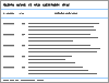
| Characteristic | Value | | Patient, n | 480 | | Age, mean (SD), year | 53.8 (14.5) | | Male, n (%) | 301 (62.7) | | Charlson score, mean (SD) | 1.6 (2) | | Preoperative hemoglobin, mean (SD), g/dL | 13.5 (1.6) | | Preoperative creatinine, mean (SD), mg/dL | 1.09 (1.5) | | Laterality, n (%) | | Right | 196 (40.8) | | Left | 260 (54.2) | | Bilateral | 24 (5.0) | | Renal stone, mean (SD), n | 1.8 (1.2) | | Diameter of largest renal stone, mean (SD), mm | 8.7 (6.1) | | Patients with preoperative double-J stent, n (%) | 271 (56.5) | | Patients with preoperative positive urine culture, n (%) | 79 (16.5) | | Duration of operation, median (range), min | 53 (15-168) |
|
|
Demographic, preoperative, and operative data of the entire cohort stratified by stone location.
|
| Characteristic | Value | | Hospital stay, median (range), day | 1 (1-14) | | Follow-up, median (range), month | 18.6 (6-161) | | Stone-free rate at long-term follow-up, n (%) | 455 (94.8) | | Stone type, n (%) | | Struvite | 10 (2.1) | | Carbonate apatite | 1 (0.2) | | Uric acid | 37 (7.7) | | Cystine | 3 (0.6) | | Calcium oxalate | 167 (34.8) | | Calcium phosphate | 2 (0.4) | | No analysis performed | 259 (54.0) | | Postoperative complication, n (%) | | Fever | 11 (2.3) | | Urosepsis | 5 (1.0) | | Perforation of renal pelvis | 0 | | Steinstrasse | 1 (0.2) | | Urinary tract infection | 11 (2.3) | | Ureteral stricture | 0 |
|
|
Postoperative complications and follow-up data following treatment of lower pole stones (n=480).
|
| Characteristic | OR | 95% CI | p-Value | | Gender (female) | 1.250 | 0.647-2.414 | 0.5 | | Age, year | 1.011 | 0.989-1.033 | 0.3 | | Renal stone, n | 0.924 | 0.709-1.203 | 0.5 | | Cumulative stone diameter, mm | 0.893 | 0.837-0.952 | 0.001 | | Ureteral stent prior to procedure | 0.511 | 0.270-0.964 | 0.03 |
|
|
Multivariable logistic regression models predicting success in RIRS for LPS below 10 mm.
|
| Characteristic | OR | 95% CI | p-Value | | Gender (female) | 1.158 | 0.711-1.883 | 0.5 | | Age, year | 0.991 | 0.974-1.008 | 0.2 | | Renal stone, n | 0.881 | 0.731-1.062 | 0.1 | | Cumulative stone diameter, mm | 0.903 | 0.867-0.941 | <0.0001 | | Ureteral stent prior to procedure | 0.515 | 0.318-0.835 | 0.007 |
|
|
Multivariable logistic regression models predicting success in RIRS for LPS above 10 mm.
|
| [1] |
Gurocak S, Kupeli B, Acar C, Tan MO, Karaoglan U, Bozkirli I. The impact of pelvicaliceal features on problematic lower pole stone clearance in different age groups. Int Urol Nephrol 2008; 40:31e7.
pmid: 17619163
|
| [2] |
Elbahnasy AM, Shalhav AL, Hoenig DM, Elashry OM, Smith DS, McDougall EM, et al. Lower caliceal stone clearance after shock wave lithotripsy or ureteroscopy: the impact of lower pole radiographic anatomy. J Urol 1998; 159:676e82.
pmid: 9474124
|
| [3] |
Sorensen CM, Chandhoke PS. Is lower pole caliceal anatomy predictive of extracorporeal shock wave lithotripsy success for primary lower pole kidney stones? J Urol 2002; 168: 2377e82.
doi: 10.1097/01.ju.0000036354.52323.c1
pmid: 12441921
|
| [4] |
Albala DM, Assimos DG, Clayman RV, Denstedt JD, Grasso M, Gutierrez-Aceves J, et al. Lower pole I: a prospective ran-domized trial of extracorporeal shock wave lithotripsy and percutaneous nephrostolithotomy for lower pole neph-rolithiasisdinitial results. J Urol 2001; 166:2072e80.
doi: 10.1016/s0022-5347(05)65508-5
pmid: 11696709
|
| [5] |
Zanetti G, Seveso M, Montanari E, Guarneri A, Del Nero A, Nespoli R, et al. Renal stone fragments following shock wave lithotripsy. J Urol 1997; 158:352e5.
pmid: 9224301
|
| [6] |
Hesse A, Br?ndle E, Wilbert D, K?hrmann KU, Alken P. Study on the prevalence and incidence of urolithiasis in Germany comparing the years 1979 vs. 2000. Eur Urol 2003; 44:709e13.
|
| [7] |
Drach GW, Dretler S, Fair W, Finlayson B, Gillenwater J, Grif?th D, et al. Report of the United States cooperative study of extracorporeal shock wave lithotripsy. J Urol 1986; 135: 1127e33.
|
| [8] |
Grasso M, Ficazzola M. Retrograde ureteropyeloscopy for lower pole caliceal calculi. J Urol 1999; 162:1904e8.
doi: 10.1016/s0022-5347(05)68065-2
pmid: 10569534
|
| [9] |
Wendt-Nordahl G, Mut T, Krombach P, Michel MS, Knoll T. Do new generation flexible ureterorenoscopes offer a higher treatment success than their predecessors? Urol Res 2010; 39: 185e8.
doi: 10.1007/s00240-010-0331-0
|
| [10] |
Ito H, Sakamaki K, Kawahara T, Terao H, Yasuda K, Kuroda S, et al. Development and internal validation of a nomogram for predicting stone-free status after flexible ureteroscopy for renal stones. BJU Int 2015; 115:446e51.
doi: 10.1111/bju.12775
pmid: 24731157
|
| [11] |
Chautard D, Bigot P, Azzouzi AR, Pichon T, Chautard D, Culty T, et al. Impact of lower pole calculi in patients un-dergoing retrograde intrarenal surgery. J Endourol 2013; 28: 141e5.
doi: 10.1089/end.2013.0515
|
| [12] |
Auge BK, Dahm P, Wu NZ, Preminger GM. Ureteroscopic management of lower-pole renal calculi: technique of calcu-lus displacement. J Endourol 2002; 15:835e8.
doi: 10.1089/089277901753205852
|
| [13] |
Türk C, Neisius A, Pet?ík A, Seitz C, Skolarikos A, Somani B, et al. European association of Urology guidelines. 2018 edi-tion. The European Association of Urology Guidelines Of?ce; 2018. Available at: http://uroweb.org/guideline/urolithiasis/. [Accessed 1 January 2020].
|
| [14] |
Goldberg H, Golomb D, Shtabholtz Y, Tapiero S, Creiderman G, Shariv A, et al. The “old” 15 mm renal stone size limit for RIRS remains a clinically signi?cant threshold size. World J Urol 2017; 35:1947e54.
doi: 10.1007/s00345-017-2075-8
pmid: 28756558
|
| [15] |
Donaldson JF, Lardas M, Scrimgeour D, Stewart F, MacLennan S, Lam TBL, et al. Systematic review and meta-analysis of the clinical effectiveness of shock wave litho-tripsy, retrograde intrarenal surgery, and percutaneous nephrolithotomy for lower-pole renal stones. Eur Urol 2015; 67:612e6.
doi: 10.1016/j.eururo.2014.09.054
pmid: 25449204
|
| [16] |
Bernardini S, Pastori J, Jacquemet B, Bailly V, Guichard G, Bernardini S, et al. Comparison of the ef?cacy and morbidity of flexible ureterorenoscopy for lower pole stones compared with other renal locations. J Endourol 2014; 28:1183e7.
doi: 10.1089/end.2014.0286
pmid: 24811281
|
| [17] |
Martin F, Hoarau N, Lebdai S, Pichon T, Chautard D, Culty T, et al. Impact of lower pole calculi in patients un-dergoing retrograde intrarenal surgery. J Endourol 2014; 28: 141e5.
doi: 10.1089/end.2013.0515
|
| [18] |
Kourambas J, Delvecchio FC, Munver R, Pichon T, Chautard D, Culty T, et al. Nitinol stone retrieval-assisted ureteroscopic management of lower pole renal calculi. Urology 2000; 56: 935e9.
pmid: 11113736
|
| [19] |
Schuster TG, Hollenbeck BK, Faerber GJ, Wolf JS. Uretero-scopic treatment of lower pole calculi: comparison of litho-tripsy in situ and after displacement. J Urol 2002; 168:43e5.
pmid: 12050489
|
| [1] |
Jirong Lu,Karthik Thandapani,Tricia Kuo,Ho Yee Tiong. Validation of laparoscopy and flexible ureteroscopy tasks in inanimate simulation training models at a large-scale conference setting[J]. Asian Journal of Urology, 2021, 8(2): 215-219. |
| [2] |
Etienne Xavier Kellera,Vincent De Conincka,Steeve Doizia,Olivier Traxer. The role of ureteroscopy for treatment of staghorn calculi: A systematic review[J]. Asian Journal of Urology, 2020, 7(2): 110-115. |
| [3] |
Jad Khaled AlSmadi,Xiaohang Li,Guohua Zeng. Use of a modified ureteral access sheath in semi-rigid ureteroscopy to treat large upper ureteral stones is associated with high stone free rates[J]. Asian Journal of Urology, 2019, 6(3): 217-221. |
| [4] |
Jennifer Bjazevic,Hassan Razvi*. Stones in pregnancy and pediatrics[J]. Asian Journal of Urology, 2018, 5(4): 223-234. |
| [5] |
Itay M. Sabler,Ioannis Katafigiotis,Ofer N. Gofrit,Mordechai Duvdevani. Present indications and techniques of percutaneous nephrolithotomy: What the future holds?[J]. Asian Journal of Urology, 2018, 5(4): 287-294. |
| [6] |
María Rodríguez-Monsalve Herrero,Steeve Doizi,Etienne Xavier Keller,Vincent De Coninck,Olivier Traxer. Retrograde intrarenal surgery: An expanding role in treatment of urolithiasis[J]. Asian Journal of Urology, 2018, 5(4): 264-273. |
| [7] |
Dong Hao,Peng Yonghan,Li Ling,Gao Xiaofeng. Prevention strategies for ureteral stricture following ureteroscopic lithotripsy[J]. Asian Journal of Urology, 2018, 5(2): 94-100. |
| [8] |
Akio Takayanagi, Atsushi Takahashi, Fumimasa Fukuta, Manabu Okada, Masahiro Matsuki, Shunsuke Sato, Teruhisa Uehara, Shuichi Kato, Yoshio Takagi. Who needs further evaluations to diagnose upper urinary tract urothelial cancers among patients with abnormal fi ndings by enhanced CT?[J]. Asian Journal of Urology, 2016, 3(1): 44-48. |
| [9] |
Zhixiang Wang, Bing Liu, Xiaofeng Gao, Yi Bao, Yang Wang, Huamao Ye, Yinghao Sun, Linhui Wang. Laparoscopic ureterolysis with simultaneous ureteroscopy and percutaneous nephroscopy for treating complex ureteral obstruction after failed endoscopic intervention: A technical report[J]. Asian Journal of Urology, 2015, 2(4): 238-243. |
| [10] |
Husain Alenezi, John D. Denstedt. Flexible ureteroscopy: Technological advancements, current indications and outcomes in the treatment of urolithiasis[J]. Asian Journal of Urology, 2015, 2(3): 133-141. |
| [11] |
Christopher Netsch, Sophie Knipper, Christian Tiburtius, Andreas J. Gross. Systematic evaluation of a holmium: yttrium-aluminumgarnet laser lithotripsy device with variable pulse peak power and pulse duration[J]. Asian Journal of Urology, 2014, 1(1): 57-61. |
|
|
|
|